India’s healthcare sector has witnessed remarkable technological advancement over the past decade. Hospitals and diagnostic centres are rapidly adopting advanced imaging systems such as MRI, CT, PET-CT, digital X-ray, mammography, and AI-assisted radiology workflows. Healthcare institutions proudly invest in cutting-edge imaging equipment to improve diagnosis and patient care.
However, a critical vulnerability exists within these advanced diagnostic environments. Medical professionals frequently overlook display calibration, which constitutes a severe patient safety issue in Indian healthcare. Facilities rigorously maintain the imaging machines but routinely neglect the monitors that display the final images.
This silent oversight directly degrades diagnostic precision. A radiologist relies entirely on the monitor to detect microscopic abnormalities. When healthcare administrators ignore display calibration, they compromise reporting confidence and negatively impact patient outcomes.
What Is Display Calibration?
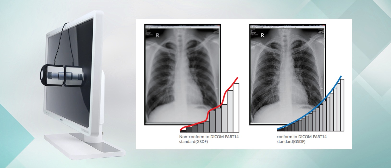
Display calibration adjusts medical monitors to DICOM standards, ensuring accurate grayscale and brightness for diagnostic imaging. In India, uncalibrated displays remain a critical patient safety risk, causing radiologists to miss subtle pathologies in mammography and chest X-rays due to degraded contrast and inconsistent image quality.
Calibration ensures that:
- Images appear consistently across workstations
- Grayscale shades are accurately displayed
- Brightness remains stable over time
- Diagnostic details remain visible
Without calibration, monitors gradually drift from their original performance levels, often without users realizing it.
How Do Advanced Imaging Systems Impact Modern Healthcare in India?
Advanced imaging systems are highly specialized diagnostic tools that capture detailed anatomical structures. These modalities generate high-resolution data that physicians use to identify diseases at early stages.
Advanced imaging systems perform the following critical functions:
- Capture precise cross-sectional views of internal organs.
- Highlight structural abnormalities in neurological and musculoskeletal systems.
- Provide functional data regarding cellular activity in oncology cases.
- Feed high-fidelity pixel data into AI algorithms for preliminary analysis.
Despite the sophisticated technology generating the data, the human eye must interpret the final visual output. The entire diagnostic process depends on the accurate visual representation of the scan. If the display alters the contrast or luminance of the image, the radiologist evaluates a flawed representation of the patient’s anatomy.
What Does Medical Display Calibration Involve?
Display calibration is the technical process of adjusting a monitor to meet specific medical imaging standards. Clinical engineers configure the brightness, contrast, grayscale response, and colour accuracy of the monitor to match the Digital Imaging and Communications in Medicine (DICOM) Part 14 standard. According to the National Electrical Manufacturers Association [NEMA, 2024], the DICOM standard uses a mathematical model called the Grayscale Standard Display Function (GSDF) to ensure that the human eye perceives consistent changes in luminance.
Display calibration ensures that monitors maintain the following parameters:
- Images appear consistently across different workstations and departments.
- Grayscale shades map accurately to the specific tissue densities captured by the scanner.
- Brightness levels remain stable over the entire lifespan of the display.
- Subtle diagnostic details remain visible in both the darkest and lightest areas of the image.
Without regular calibration, a monitor gradually drifts from its original baseline. The liquid crystal display (LCD) components degrade over time, leading to a loss of peak luminance. Users rarely notice this gradual degradation until the monitor fails to display critical clinical information.
How Do Uncalibrated Displays Compromise Patient Safety?
Uncalibrated monitors introduce multiple points of failure into the diagnostic workflow. These failures directly compromise patient safety by increasing the likelihood of diagnostic errors.
Why Do Missed or Delayed Diagnoses Occur?
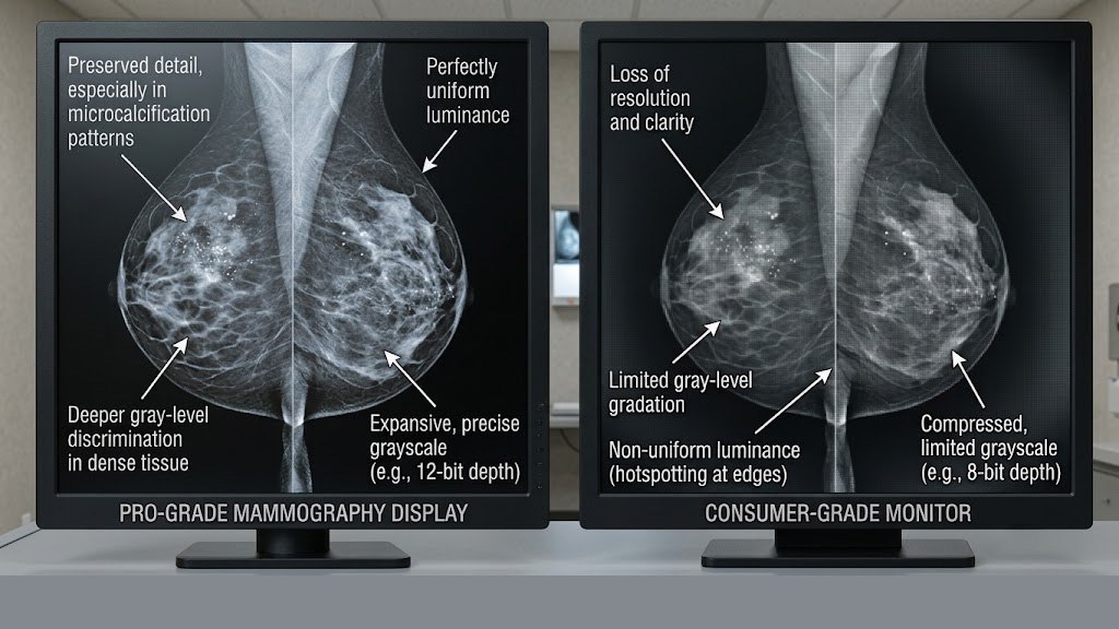
Poor luminance and inadequate contrast ratios hide subtle clinical findings. A microcalcification in breast tissue or a small nodule in the lung may blend into the background of a degraded monitor.
Missed diagnoses occur most frequently in the following clinical scenarios:
- Mammography: Breast imaging requires exceptionally high luminance (often exceeding 400 nits) to differentiate dense tissue.
- Chest imaging: Radiologists must detect faint ground-glass opacities that indicate early-stage pulmonary conditions.
- Neuroimaging: Physicians require precise grayscale separation to identify acute ischemic strokes.
- Oncology follow-ups: Clinicians must measure minor changes in tumour size across multiple scans taken months apart.
When a radiologist misses a lesion due to a poor display, the patient experiences a delayed diagnosis. This delay directly reduces treatment efficacy and survival rates.
What Causes Inconsistent Image Interpretation?
Inconsistent image interpretation happens when uncalibrated monitors display the same data differently. A scan may look perfectly normal on one workstation but show suspicious shadows on another.
Without DICOM-compliant calibration, facilities experience the following issues:
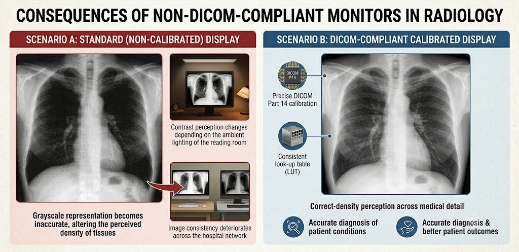
- Grayscale representation becomes inaccurate, altering the perceived density of tissues.
- Contrast perception varies with the ambient lighting in the reading room.
- Image consistency deteriorates across the hospital network.
Consequently, two radiologists may provide conflicting reports for the same patient simply because they view the images on monitors with different calibration statuses.
How Does Monitor Quality Affect Radiologist Fatigue?
Radiologists spend intensive hours reading hundreds of complex images every day. Uncalibrated or consumer-grade monitors exhibit poor brightness stability, uneven backlighting, and subtle flickering.
Visual fatigue causes the following operational detriments:
- Reduces the radiologist’s concentration during long shifts.
- Slows down the overall reporting turnaround time.
- Increases the frequency of perceptual reading errors.
- Lowers the productivity of the entire diagnostic department.
A high-performance diagnostic monitor mitigates eye strain and maintains reading efficiency, thereby protecting the radiologist’s cognitive bandwidth.
What Are the Compliance and Standards for Medical Imaging Displays?
Healthcare standards boards strongly emphasize image quality assurance. In India, the National Accreditation Board for Hospitals & Healthcare Providers [NABH, 2025] requires medical imaging services to monitor key performance indicators, including the rate of radiology reporting errors.
Using non-medical-grade displays creates significant compliance concerns during institutional audits. Medical-grade monitors are specialized hardware units that manufacturers build specifically to meet rigorous clinical guidelines.
Medical-grade monitors comply with the following requirements:
- Maintain DICOM Part 14 GSDF compliance automatically via built-in front sensors.
- Guarantee consistent luminance output over thousands of hours of use.
- Support comprehensive quality assurance (QA) software that logs performance data.
Hospitals that ignore display quality expose their organizations to substantial legal and operational risks, particularly when diagnostic errors lead to malpractice claims.
Why Does Display Calibration Remain Overlooked in India?
Display calibration remains a neglected priority in India for several systemic reasons. Firstly, the issue is invisible. Because monitor degradation happens slowly, radiologists adapt to the dimming screen without realizing the loss of contrast.
Secondly, hospital administrators often underestimate the impact of visualization hardware on patient outcomes. Procurement departments enthusiastically approve costly budgets for new MRI machines but attempt to save funds by purchasing consumer-grade monitors for the radiology reading rooms. This misallocation of resources breaks the imaging chain at its most crucial link.
How Can Indian Healthcare Institutions Improve Display Calibration?
Healthcare leaders must treat the diagnostic monitor as a medical device rather than a generic computer accessory. Correcting the current oversight requires a structured approach to quality assurance.
Institutions must implement the following solutions:
- Implement regular calibration schedules using centralized quality control software that alerts administrators when a display falls out of compliance.
- Invest exclusively in medical-grade monitors for primary diagnostic reading, ensuring the hardware contains built-in calibration sensors.
- Provide comprehensive training for healthcare professionals regarding the impact of ambient light and display settings on diagnostic accuracy.
- Encourage regulatory bodies to enforce stricter guidelines specifically targeting display monitor quality assurance during facility inspections.
What Are the Next Steps for Diagnostic Accuracy?
Display calibration plays a foundational role in patient safety. While it remains largely invisible and gradual in its degradation, its impact on diagnostic precision is absolute. Indian healthcare institutions cannot afford to compromise the final stage of the imaging chain. The data captured by an advanced scanner holds no value if the display cannot present it accurately to the diagnosing physician.
As India modernizes its medical infrastructure, display quality and calibration must transition from an optional administrative task to a mandatory clinical priority. Healthcare administrators and radiology directors must audit their current reading environments and upgrade their visualization tools. Because in medical imaging, what the radiologist sees determines the treatment the patient receives.
Frequently Asked Questions
What is the difference between a medical-grade monitor and a consumer monitor?
Medical-grade monitors contain specialized sensors that continuously measure and adjust luminance to maintain DICOM Part 14 compliance. They provide higher peak brightness, longer lifespans, and uniform backlighting. Choose a medical-grade monitor if you require diagnostic accuracy for clinical decision-making, as consumer monitors cannot reliably display the subtle grayscale differences necessary for identifying pathologies.
How often should hospitals calibrate diagnostic displays?
According to international radiology guidelines, hospitals should perform automated quality control checks daily and conduct full physical calibrations at least annually. Facilities should utilize centralized quality assurance software to track monitor performance continuously and flag any displays that drift beyond acceptable luminance parameters.
Which imaging modalities require the highest display calibration standards?
Mammography requires the strictest calibration standards due to the extremely low contrast nature of breast tissue anomalies. Displays used for mammography must sustain high luminance levels (typically 400 to 1000 nits) to reveal microcalcifications. Choose a specialized 5-megapixel (or higher) medical display if your department interprets digital breast tomosynthesis or screening mammograms.
Conclusion
Display calibration remains one of India’s most overlooked patient safety issues because it is invisible, gradual, and often underestimated. Yet its impact on diagnostic accuracy can be enormous.
Healthcare institutions cannot afford to ignore the final stage of the imaging chain — image visualization itself.
As India continues modernizing its healthcare infrastructure, display quality and calibration must become a standard clinical priority rather than an afterthought. Because in medical imaging, what the radiologist sees determines what the patient receives.