
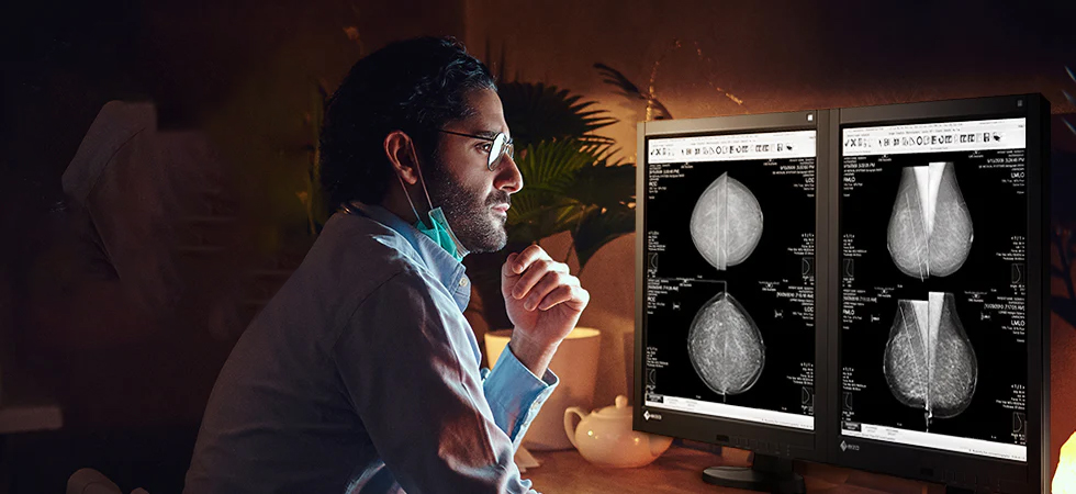
Every year, breast cancer claims the lives of hundreds of thousands of women worldwide. Yet when doctors detect it early — at stage I — survival rates soar above 99%. That number tells a powerful story: the sooner clinicians find cancer, the better the outcome. And at the center of early detection sits one critical, often overlooked piece of technology: the mammography display.
Radiologists examine mammograms on medical-grade monitors for hours each day. What they see — and crucially, what they can distinguish — shapes every diagnosis. High-resolution mammography monitors do not simply show clearer images. They enable radiologists to find cancers that standard monitors miss entirely.
Understanding Mammography: What Radiologists Look For
Digital mammography produces highly detailed grayscale images of breast tissue. Radiologists examine these images for three key abnormalities: microcalcifications (tiny calcium deposits that can signal early ductal carcinoma in situ), masses or lumps within the tissue, and architectural distortions — subtle warping of normal breast structures that often indicates an invasive tumor.
Each of these findings demands extraordinary precision. Microcalcifications, for instance, measure less than 0.5 millimeters. A radiologist must detect a cluster of specks smaller than a grain of salt against a complex, layered background. Any loss of image fidelity — blurred edges, insufficient contrast, washed-out grays — can make these findings invisible.
This is where display quality stops being a technical specification and starts being a matter of patient survival.
What Makes a Mammography Display High-Resolution?
Not every computer monitor qualifies as a mammography display. Diagnostic-grade mammography monitors meet a demanding set of technical benchmarks that consumer displays simply cannot match.
Pixel density leads the list. Standard HD monitors deliver 1–2 megapixels. Diagnostic mammography displays operate at 5 megapixels or higher — enough to render the fine-grained structure of breast tissue without losing detail. Some leading systems now offer 10–12 megapixel panels for full-field digital mammography.
Luminance levels matter just as much. Medical displays must achieve peak brightness of 500–600 cd/m² (candelas per square meter) to ensure radiologists can distinguish subtle tissue density differences. Consumer monitors typically reach only 250–300 cd/m².
DICOM (Digital Imaging and Communications in Medicine) compliance ensures the display renders grayscale values in a standardized, clinically validated way. Without DICOM calibration, the same image can look dramatically different across two monitors — a dangerous inconsistency in a diagnostic setting.
“A 5-megapixel diagnostic display renders up to five times more pixel information than a standard HD monitor — each additional pixel is an opportunity to see something critical.”
How High-Resolution Displays Improve Early Detection
The link between display quality and diagnostic accuracy is direct and well-established. High-resolution mammography monitors improve detection in several concrete ways.
They reveal microcalcifications earlier. Because high-resolution displays render fine detail with greater fidelity, radiologists can identify clusters of microcalcifications at a smaller size and earlier stage — before they develop into invasive cancers. Studies have shown that pixel pitch (the distance between pixels) directly affects detection sensitivity for small calcifications.
They reduce eye strain and cognitive fatigue. Radiologists read dozens to hundreds of mammograms per shift. Low-quality displays force the eye to work harder, amplifying fatigue. High-resolution, well-calibrated monitors reduce visual strain, helping radiologists maintain focus and accuracy throughout a long workday.
They improve confidence in ambiguous findings. In borderline cases, a radiologist decides between recalling a patient for additional imaging or clearing them. Better display quality gives clinicians more information to work with, reducing both false positives (unnecessary anxiety and procedures) and false negatives (missed cancers).
They support 3D tomosynthesis review. Digital breast tomosynthesis (DBT), or 3D mammography, generates hundreds of thin image slices per study. Reviewing these effectively requires displays that can render fine detail consistently across every slice. High-resolution monitors make this workflow faster and more accurate.
The Clinical Evidence
Research consistently confirms the link between display quality and diagnostic performance. Studies examining radiologist performance across different monitor types show that higher-resolution displays improve sensitivity for detecting small masses and microcalcification clusters — the findings most critical to early-stage diagnosis.
One consistent finding across the literature: radiologists using sub-optimal displays miss more cancers at smaller sizes. They also call back patients more frequently for follow-up imaging — driving up costs and patient anxiety — because they cannot confidently interpret what they see.
Radiology departments that invest in high-quality diagnostic displays report improvements in radiologist confidence, reductions in unnecessary recall rates, and stronger early-stage detection outcomes.
“The difference between a diagnostic-grade display and a standard monitor is not cosmetic. It is the difference between seeing a 2mm lesion and missing it entirely.”
Regulatory Standards: What the Guidelines Require
Regulators and professional bodies recognize the critical role displays play in mammography accuracy. The Mammography Quality Standards Act (MQSA) in the United States sets requirements for mammography equipment, including display systems used for diagnostic interpretation.
The American College of Radiology (ACR) and the American Association of Physicists in Medicine (AAPM) publish guidelines specifying luminance ratios, pixel resolution, and calibration protocols for mammography workstations. Facilities must conduct regular quality control checks — including display luminance testing — to maintain accreditation.
These standards exist for a reason: display quality is not optional in breast imaging. It is a regulatory requirement and a clinical obligation.
The Cost-Benefit Argument for Investing in Better Displays
Healthcare administrators sometimes hesitate at the price of premium diagnostic displays. A high-resolution mammography monitor can cost several times more than a consumer-grade screen. But this comparison misses the full financial picture.
A missed breast cancer at stage I costs far more to treat when it progresses to stage III or IV. The difference in treatment costs between early and late-stage breast cancer runs into tens of thousands of dollars per patient. Add litigation risk from missed diagnoses, and the economic case for investing in diagnostic-grade displays becomes compelling.
Equally important: every mammogram a radiologist reads with superior display quality is an opportunity to find cancer sooner, treat it more effectively, and help a patient live longer. That outcome does not appear on a balance sheet, but it drives the entire mission of breast cancer screening.
The Future: AI, Tomosynthesis, and the Next Generation of Displays
Artificial intelligence is transforming mammography interpretation. AI-assisted detection tools analyze mammogram images and flag suspicious regions for radiologist review. But AI algorithms depend on the same image data the radiologist sees — and they perform best when displays render that data with maximum fidelity.
As 3D tomosynthesis becomes the standard of care at more facilities, display demands will only increase. Reviewing a full DBT study involves hundreds of images per patient. Displays must handle high pixel counts, consistent calibration across large panels, and ergonomic designs that reduce fatigue during extended reading sessions.
Manufacturers are already developing the next generation of diagnostic displays — higher pixel densities, improved HDR (high dynamic range) capabilities, and integrated AI overlays that highlight areas of interest without obscuring underlying detail. The radiologist of tomorrow will read mammograms on displays that show more, reveal more, and catch more — if healthcare institutions make the investment today.
Conclusion: Display Quality Is Patient Safety
Early breast cancer detection saves lives. That is not a slogan — it is a statistical reality backed by decades of clinical data. But detection depends on the complete diagnostic chain: a properly positioned image, a skilled radiologist, and a display capable of showing every critical detail.
High-resolution mammography displays form the final link in that chain. When facilities invest in diagnostic-grade monitors, they give radiologists the tools to find cancers earlier, interpret images with greater confidence, and deliver better outcomes for patients.
The technology exists. The evidence is clear. The choice to equip imaging departments with high-resolution mammography displays is not a technical upgrade — it is a commitment to the patients who walk through the door hoping for a clear answer.
