Walk into any hospital’s radiology department and you’ll see rows of monitors that all look, at first glance, remarkably similar. Dark bezels, matte screens, grayscale test patterns humming quietly in the background. But ask a radiologist which screen they’d trust to report a chest CT, and which one they’d hand to a surgeon reviewing prior scans before a procedure, and you’ll get two very different answers. The distinction between a diagnostic display and a clinical review display isn’t cosmetic; it’s a matter of patient safety, workflow efficiency, and regulatory compliance.
Hospitals and imaging centers frequently conflate the two, either because procurement teams assume “a medical monitor is a medical monitor,” or because vendors don’t always explain the engineering differences clearly. This article breaks down exactly what separates these two categories of medical monitors, where each belongs in a modern radiology workflow, and how to avoid the costly mistake of using the wrong display for the wrong task.
What Is a Diagnostic Display?
A diagnostic display is a purpose-built radiology monitor engineered specifically for primary image interpretation the act of a radiologist reading a study and rendering a formal diagnosis. These are the monitors mounted in reading rooms, and they are built around three non-negotiable pillars: resolution, luminance, and calibration.
Resolution and pixel density.Diagnostic displays typically range from 3MP up to 12MP, depending on the modality. A 3MP or 5MP grayscale monitor is standard for general radiography and CT/MRI review, while mammography and tomosynthesis studies demand 5MP color or higher, and dual-head 12MP setups are common for side-by-side comparison of high-resolution mammographic images. The pixel pitch on these panels is tight enough that a radiologist can detect a microcalcification cluster or a subtle nodule without digital zoom artifacts degrading the image.
Luminance and grayscale rendering. Diagnostic monitors are built to sustain high peak luminance generally 400 to 1,000 cd/m² because subtle density differences in a chest X-ray or a lung window CT slice depend entirely on how accurately the panel reproduces shades of gray. A monitor that can’t hold consistent luminance across its full grayscale range will hide pathology in the shadows or blow it out in the highlights.
DICOM GSDF calibration. Every diagnostic display must be calibrated to the DICOM Part 14 Grayscale Standard Display Function (GSDF), which ensures that the same pixel value produces the same perceived brightness step on every monitor in the department, regardless of manufacturer or age. This is what allows two radiologists reading the same study on two different diagnostic monitors to see, functionally, the same image. Diagnostic displays typically include built-in front sensors or require periodic calibration through software like MediCal QAWeb, so drift in brightness or color temperature is caught before it affects a read.
In short: a diagnostic display exists to answer one question with total confidence: is this finding real, and how significant is it?
What Is a Clinical Review Display?
A clinical review display, by contrast, is built for secondary viewing the review of already-interpreted images by referring physicians, surgeons, ward consultants, ICU staff, or clinicians in an OPD setting who need to see the scan, understand the context, and make a treatment decision, but who are not rendering the primary radiological report.
These displays are typically lower resolution, often 1MP to 3MP and use standard color LCD panels rather than the specialized grayscale or dual-driver panels found in diagnostic monitors. Luminance requirements are more relaxed, generally in the 250–350 cd/m² range, since the use case is contextual review rather than pixel-level pathology detection. Calibration, when applied, is usually lighter-touch, often limited to basic brightness and contrast standardization rather than full DICOM GSDF compliance.
Clinical review displays are the workhorses of the hospital outside the reading room: nursing stations, surgeon’s offices, ICU consoles, telemedicine carts, and OPD consultation rooms. They’re connected to PACS or a viewer like OsiriX, letting any authorized clinician pull up a patient’s imaging history quickly, without needing reading-room-grade hardware.
Key Technical Differences at a Glance
Parameter
Diagnostic Display
Clinical Review Display
Typical Resolution
3MP–12MP
1MP–3MP
Panel Type
Grayscale or medical-grade color LCD
Standard color LCD
Peak Luminance
400–1,000 cd/m²
250–350 cd/m²
DICOM GSDF Calibration
Mandatory, front-sensor or software-driven
Optional / basic
Primary Use
First-read diagnosis by radiologist
Secondary review by clinicians
Location
Reading room
Ward, OPD, surgeon’s office, ICU
Regulatory Status
Cleared for primary diagnosis
Not intended for primary diagnosis
The gap isn’t just about specs on a datasheet it changes how the two devices behave under real clinical conditions. A diagnostic monitor holds its calibration under continuous use across long reporting shifts; a clinical review display is optimized for cost-efficiency and everyday usability rather than sustained diagnostic-grade precision.
Why Ambient Light and Panel Technology Matter
Reading rooms are deliberately kept dim, with controlled, indirect lighting, because ambient light reflecting off a monitor’s surface reduces perceived contrast and can mask low-density findings. Diagnostic displays are engineered with anti-glare, anti-reflective coatings and matte finishes to perform reliably in these controlled environments.
Clinical review displays, on the other hand, are used in brightly lit wards, nursing stations, and consultation rooms where ambient light is uncontrolled. Manufacturers compensate with higher baseline brightness and glossier or semi-glossy coatings suited for general visibility rather than diagnostic contrast fidelity, a sensible trade-off for a device that isn’t being used to detect a 2mm nodule.
Regulatory and Compliance Considerations
This is where the distinction stops being a matter of preference and becomes a matter of compliance. Regulatory bodies distinguish clearly between displays intended for primary diagnosis and those intended for review only. In most jurisdictions, only monitors that meet DICOM GSDF calibration standards and pass photometric testing protocols such as AAPM TG18 are cleared for primary radiological reporting.
Using a clinical review display or worse, an uncalibrated consumer-grade monitor to render a primary diagnosis isn’t just poor practice; it can constitute a compliance violation and introduces real diagnostic risk. Regulatory and accreditation bodies increasingly expect hospitals to document their calibration and QA processes for diagnostic displays, which is why routine monitor calibration services and QA software are now considered essential infrastructure, not optional add-ons.
Common Mistakes Hospitals Make
Having worked across imaging deployments in Indian hospital procurement environments, a few recurring mistakes stand out:
Buying on resolution alone. A 5MP monitor without proper GSDF calibration is not automatically “diagnostic grade.” Calibration and luminance stability matter as much as pixel count.
Deploying diagnostic displays in non-reading-room settings to save on a second purchase this wastes budget on hardware that’s overspecified for ward-level review needs.
Deploying clinical review displays in the reading room to cut upfront costs, which risks missed findings, radiologist eye strain, and non-compliance with diagnostic imaging standards.
Skipping periodic recalibration. Even a properly specified diagnostic monitor drifts in luminance and grayscale accuracy over months of use. Without a calibration schedule, a hospital can be reading on a monitor that no longer meets the standard it was purchased to meet.
Ignoring PACS integration compatibility. A display is only as useful as the software driving it — resolution and calibration profiles should be matched against the hospital’s PACS software and workstation configuration before purchase.
How to Choose the Right Display for Your Facility
When specifying medical monitors for a new department or an upgrade cycle, work through these questions in order:
Who is the primary user? Radiologist rendering first reads → diagnostic display. Referring physician, surgeon, or nurse reviewing prior studies → clinical review display.
What modality dominates the workload? Mammography and tomosynthesis push you toward higher-resolution color diagnostic displays; general radiography and CT/MRI reporting are typically well served by 3MP–5MP grayscale diagnostic monitors.
What’s the ambient lighting environment? Controlled reading room versus brightly lit ward changes both the panel coating and the luminance specification you need.
What’s your calibration and QA capacity? Diagnostic displays require an ongoing calibration program — factor in software like MediCal QAWeb and a maintenance schedule, not just the upfront hardware cost.
What does your PACS and workstation software support? Confirm compatibility before locking in a display resolution or panel type.
Conclusion
Diagnostic and clinical review displays serve two distinct, equally important roles in the modern radiology workflow — one exists to support the radiologist’s primary act of interpretation with clinical-grade precision, and the other exists to extend that imaging data efficiently across the rest of the care team. Treating them as interchangeable, or choosing based on price alone, puts both diagnostic accuracy and regulatory compliance at risk.
Proscreen Technologies works with hospitals and diagnostic centers across India to specify, supply, and calibrate the right mix of diagnostic displays, clinical review displays, and mammography displays for their exact workflow backed by PACS software integration, AI-assisted radiology tools, and ongoing monitor calibration services to keep every screen in the department performing to standard, year after year.
Walk into most operating rooms built before 2015, and you’ll find the same setup: a hardware matrix switch bolted into an equipment rack, feeding a handful of displays through dedicated coax or SDI cables. This design served hospitals well for years. But surgical video has changed faster than the hardware built to carry it, and matrix switches are struggling to keep up.
Today’s ORs push 4K endoscopy feeds, C-arm imaging, patient vitals, and room cameras through the same infrastructure — often to multiple displays inside the room and outside it. Surgeons now expect to pull up a scope feed next to a PACS image on the same screen, stream a procedure to a lecture hall, or loop in a remote specialist mid-case. Matrix switches were never built for that level of demand, and hospitals are feeling the strain.
This is why more facilities are replacing matrix switches with IP-based video integration and understanding the difference matters before you plan your next OR build or retrofit.
How a Traditional Matrix Switch Works
A matrix switch is a physical hardware box with a fixed grid of inputs and outputs. Each video source — a scope, a camera, a monitor feed — runs through its own dedicated cable straight into the switch. The switch then routes each input to one or more outputs based on a hardwired configuration.
This model creates four problems that grow worse as your OR gets busier:
It hits a hard capacity ceiling. Every matrix switch supports a fixed number of inputs and outputs. Once you fill those slots, you can’t add a new camera or display without buying a bigger switch or a second unit.
It demands heavy, source-specific cabling. Each device needs its own dedicated run back to the switch. In a retrofit, that means opening ceilings and walls to pull new coax or SDI cable for every additional source, a costly, disruptive process.
It locks you into a fixed video standard. Matrix switches are built around the resolution and format available at the time of purchase. When your hospital moves to 4K endoscopy or a new imaging format arrives, you’re often looking at a full hardware replacement, not an upgrade.
It can’t reach beyond the room. Sending a live feed to a classroom, a control room, or a remote specialist typically requires separate dedicated infrastructure that a matrix switch was never designed to support.
How IP-Based OR Video Integration Works
An IP-based system removes the fixed hardware grid entirely. Instead of wiring each source directly to a switch, every camera, scope, and display connects to the hospital’s standard IP network through an encoder or decoder. Routing happens in software — any source can reach any display, anywhere on the network, without a single cable change.
Our Operating Room Integration system runs on this exact model, built on SDVoE (Software-Defined Video over Ethernet) hardware paired with the iVideo OR management platform. Here’s what that architecture delivers in practice:
It streams uncompressed 4K60 video with near-zero latency. SDVoE moves video at 4K60 (3840×2160 @ 60fps) with sub-frame latency — imperceptible during live surgery, where hand-eye coordination depends on real-time feedback. Compression artifacts, which can obscure fine tissue detail, never enter the signal chain.
It scales without new cabling. Adding a source or display means adding one SDVoE encoder or decoder to the existing hospital LAN not rewiring the room or upgrading a switch. The same 10GbE Ethernet infrastructure that already runs through most hospitals carries the video.
It future-proofs the investment. Because routing and control live in software, new devices, resolutions, and workflows can be integrated as they emerge, rather than forcing a hardware refresh every few years.
It extends past the OR walls. A feed can reach a lecture hall, a control room, or a remote specialist’s screen using the same network — no dedicated point-to-point run required. Paired with a platform likemedVC, surgeons can stream a live case, record it, or bring in a remote consultant for real-time collaboration during the procedure itself.
It plugs directly into hospital IT tools. Since every encoder and decoder is a standard network device, your IT team can manage the system with familiar tools VLAN isolation, port monitoring, remote firmware updates, and centralized diagnostics instead of relying on a proprietary AV vendor for every change.
IP-Based OR Video vs. Traditional Matrix Switch: A Direct Comparison
Factor
Traditional Matrix Switch
IP-Based System (Proscreen)
Video quality
HD (1080p) maximum
4K60 (3840×2160)
Routing
Fixed hardware connections
Any source to any display, via software
Scalability
Requires a hardware swap to expand
Add encoders/decoders to the existing network
Cabling
Dedicated coax or SDI per source
Standard 1GbE / 10GbE Ethernet
Remote access
Not supported
Built-in remote monitoring and streaming
DICOM documentation
Manual transfer
Automated DICOM export via iVideoOR
Future upgrades
Hardware replacement
Software-driven, incremental
When a Matrix Switch Might Still Work
A matrix switch can still make sense for a single, simple OR with a small, unchanging list of sources and no plans to expand, stream, or connect remotely. If your hospital runs one theatre with two cameras and two displays and never intends to grow, the upfront simplicity of a matrix switch is hard to beat.
That said, few hospitals stay static for long. The moment you add a second OR, plan a hybrid suite, launch a teaching program, or want to document procedures for compliance, the limits of matrix switching show up fast — usually as an unplanned capital expense.
Why Hospitals Are Making the Switch Now
Surgical video demand is only climbing. Minimally invasive and robotic procedures generate more video per case, hybrid ORs need to route imaging alongside live surgical feeds, and hospitals increasingly want to document, teach, and consult remotely without leaving the sterile field. Every one of these trends favors a network-based architecture over a fixed hardware grid.
Proscreen has deployed IP-based OR integration systems across NABH-accredited hospitals and government medical colleges in Delhi NCR, Mumbai, Bengaluru, and Chennai — retrofitting existing theatres and building hybrid ORs from the ground up, often without running new fiber or replacing existing Ethernet infrastructure. The same architecture also underpins Proscreen’s Mobile OR Integration solutions, which bring 4K streaming, near-zero-latency routing, and remote consultation to surgical setups outside the traditional hospital theatre. High-fidelity output depends on the display too — Proscreen’s surgical monitors are built to render that 4K 60 signal without losing the detail the camera captured.
Making the Right Call for Your OR
Choosing between a matrix switch and an IP-based system isn’t only a technical decision — it determines how easily your OR can grow over the next five to ten years, what a retrofit will cost when your needs change, and how well your surgical teams can collaborate beyond the four walls of the room.
If you’re planning a new OR build, a hybrid suite, or a retrofit of an aging matrix-based system, it’s worth mapping out where your hospital is headed before you commit to either architecture.
For surgeons performing minimally invasive procedures, the primary interface is no longer the patient’s anatomy directly; it is the digital visualization on the surgical display. This Surgical Display acts as the critical conduit for visual information, requiring uncompromising fidelity in depth perception, tissue contrast, and color reproduction to guide instrumentation with millimeter precision. Given that the display is essentially the surgeon’s eye in the operating theater, selecting the right technology transcends basic specifications.
It requires a deep understanding of clinical requirements, regulatory standards, and operational reliability. This guide provides a rigorous framework for evaluating surgical display monitors, ensuring your procurement decisions are driven by clinical performance rather than spec-sheet marketing.
This guide walks through what actually matters when evaluating surgical monitors for the OR — the technical factors, the practical ones, and the mistakes that trip up even experienced procurement teams.
Why Surgical Monitors Aren’t Just “Medical-Grade TVs”
It’s tempting to think of a surgical monitor as a hospital-grade version of a high-end consumer display. It isn’t. The bar is set considerably higher on several fronts at once.
Color accuracy has to be clinically reliable, not just visually pleasing — the difference between healthy and compromised tissue can hinge on subtle color shifts that a consumer panel would happily smooth over or exaggerate. Grayscale performance matters just as much, particularly for imaging-heavy procedures.
Then there’s regulation. Surgical displays need to meet FDA clearance requirements, CE marking where applicable, and DICOM Part 14 grayscale standards — none of which apply to the TV in your living room. And unlike a screen you glance at occasionally, these monitors run for hours on end in a sterile environment, get cleaned constantly with harsh disinfectants, and simply aren’t allowed to fail mid-procedure.
What to Actually Look For
Resolution: 4K, 8K, or Is Full HD Still Fine?
Full HD hasn’t disappeared from the OR, but for most modern procedures, 4K is now the practical baseline — it gives surgeons the fine detail they need for laparoscopic and endoscopic work without pushing cost or file sizes to unreasonable levels.
8K is where things get more situational. It has real value in ultra-high-precision fields like microsurgery or certain robotic-assisted procedures, where every extra pixel of detail translates to better visualization. But for general surgery, 8K often adds expense without adding much the surgeon can actually use. Worth asking: does this procedure genuinely benefit from the extra resolution, or is 4K already doing the job?
Screen Size and How It Fits the Room
Bigger isn’t automatically better here — size needs to match both the procedure and the physical layout of the OR. A 55-inch display makes sense as a central visualization hub in a hybrid OR where the whole team needs a clear view. But mount that same monitor too close to a surgeon working at short range, and it becomes more distraction than asset. Viewing distance, room size, and how many people need a clear sightline should all factor into the decision — not just “go as large as the budget allows.”
Color Accuracy and Calibration
This is arguably the single most clinically important factor on the list. A monitor that renders tissue color even slightly off can affect a surgeon’s ability to distinguish healthy tissue from something that needs attention. Look for displays with consistent, verifiable calibration — not just a spec sheet claim, but a track record of holding that calibration over the monitor’s working life, since panels do drift over time. Regular professional monitor calibration services following DICOM standards are what keep that accuracy from slipping between checkups.
Brightness and Contrast
OR lighting is bright, and displays need to hold up against it. Insufficient brightness or a weak contrast ratio makes fine detail harder to distinguish, especially in dimmer areas of the image — shadows, folds, tissue depth. A strong contrast ratio isn’t a nice-to-have; it’s what keeps detail visible instead of washed out.
LED vs. OLED
Both have a place, and the right choice depends on priorities. LED panels tend to be more affordable and durable over long duty cycles, making them a solid default for general OR use. OLED offers superior contrast and color depth — genuinely striking image quality — but usually at a higher cost and, in some cases, a shorter practical lifespan under continuous use. There’s no universally “better” option here, just a tradeoff worth weighing against the procedures the monitor will primarily support.
Response Time and Latency
For robotic-assisted and minimally invasive surgery, latency isn’t a minor technical detail — it’s a safety issue. Any lag between the surgeon’s instrument movement and what appears on screen introduces risk, particularly in procedures where millimeter-level precision matters. Low latency and fast response times should be treated as non-negotiable for these use cases, not just a preference.
Built for Sterility
Surgical monitors live in an environment that would destroy a standard commercial display fairly quickly. Sealed enclosures that resist fluid ingress, antimicrobial surfaces, and housings that can withstand repeated disinfection without degrading are all essential. This is as much about infection control as it is about protecting the hardware investment.
Connectivity and OR Integration
A great display that can’t talk to the rest of the OR’s equipment isn’t much use. Compatibility with endoscopy towers, PACS software, and broader operating room integration setups should be checked early in the evaluation — not discovered as a problem after purchase. The monitor needs to fit into an existing ecosystem, not force a workaround. For facilities that move between sites or need flexibility beyond a fixed OR, it’s worth evaluating mobile OR integration options as well.
Regulatory Compliance
Non-negotiable, full stop. Confirm FDA clearance, DICOM Part 14 compliance for grayscale accuracy, and adherence to IEC 60601 electrical safety standards before anything else is considered. A monitor that fails on compliance isn’t a candidate, regardless of how good its specs look otherwise.
Durability and Expected Lifespan
Look past the purchase price to mean time between failures (MTBF), warranty terms, and the vendor’s actual support responsiveness. A cheaper monitor that needs replacing in three years, or that leaves the OR waiting on slow support during a failure, often costs more in the long run than a pricier, more reliable option.
Matching the Monitor to the Specialty
Different procedures put different demands on a display:
Laparoscopic and general surgery prioritize low latency and strong color accuracy for navigating internal anatomy with precision.
Ophthalmology — needs exceptional fine-detail resolution and color fidelity, given the scale of the structures involved.
Cardiovascular and hybrid OR settings require displays that can handle multiple simultaneous imaging feeds, since these procedures often blend surgical and interventional radiology needs. High-end Barco medical monitors, which we offer, are a common fit here, given their reputation for multi-feed handling and image consistency.
Robotic-assisted surgery — makes latency and 4K/8K clarity essential, since the surgeon’s entire spatial awareness runs through the screen. Teaching hospitals running these procedures increasingly pair displays with surgical collaboration tools like medVC for real-time remote consultation and training.
There’s rarely a single monitor that’s ideal across all of these — many hospitals end up standardizing on a core model for general use while sourcing specialty displays for high-precision departments.
Budget vs. Long-Term Value
The lowest sticker price isn’t the same as the lowest total cost. Factor in expected lifespan, how often the panel will need recalibration, likely repair frequency, and the vendor’s support terms. A monitor that costs more upfront but lasts longer and performs more consistently often works out cheaper — and safer — over its full service life. Procurement decisions made purely on initial price tend to get revisited sooner than anyone would like.
Mistakes Worth Avoiding
A few patterns show up again and again in OR equipment purchases:
Chasing resolution alone. A 4K panel with poor color calibration will underperform a well-calibrated Full HD display in real clinical use. Resolution is one variable, not the whole equation.
Skipping the integration check. A monitor that doesn’t talk cleanly to existing endoscopy or PACS systems creates ongoing friction — sometimes expensive friction — that’s entirely avoidable with an integration check before purchase.
Underestimating support. When a display fails mid-procedure, a vendor’s response time matters as much as the hardware itself. Support terms deserve as much scrutiny as the spec sheet.
Quick-Reference Checklist
Resolution matched to procedure type (4K standard, 8K for high-precision cases)
Screen size appropriate to room layout and viewing distance
Verified, stable color calibration
Strong brightness and contrast performance
Panel type (LED/OLED) matched to use case and budget
Low latency, particularly for robotic or minimally invasive procedures
Sealed, antimicrobial housing rated for repeated disinfection
Confirmed compatibility with existing OR integration systems
FDA, DICOM Part 14, and IEC 60601 compliance verified
Clear understanding of MTBF, warranty, and support responsiveness
Frequently Asked Questions
What resolution is best for a surgical monitor? 4K is the practical standard for most procedures today. 8K adds value mainly in microsurgery and select robotic-assisted cases where extreme fine detail matters.
Do surgical monitors need to be DICOM compliant? Yes, particularly for imaging-heavy procedures. DICOM Part 14 grayscale compliance, along with FDA clearance and IEC 60601 safety standards, are baseline requirements, not optional extras.
How often should a surgical monitor be calibrated? This varies by manufacturer and usage intensity, but regular scheduled calibration is essential to prevent color and grayscale drift over time. Facilities typically rely on a dedicated calibration service rather than one-time factory settings.
LED or OLED — which is better for the OR? Neither is universally better. LED tends to offer durability and lower cost for general OR use, while OLED delivers superior contrast and color depth at a higher price point and, in some cases, shorter lifespan under continuous use.
Every year, breast cancer claims the lives of hundreds of thousands of women worldwide. Yet when doctors detect it early — at stage I — survival rates soar above 99%. That number tells a powerful story: the sooner clinicians find cancer, the better the outcome. And at the center of early detection sits one critical, often overlooked piece of technology: the mammography display.
Radiologists examine mammograms on medical-grade monitors for hours each day. What they see — and crucially, what they can distinguish — shapes every diagnosis. High-resolution mammography monitors do not simply show clearer images. They enable radiologists to find cancers that standard monitors miss entirely.
Understanding Mammography: What Radiologists Look For
Digital mammography produces highly detailed grayscale images of breast tissue. Radiologists examine these images for three key abnormalities: microcalcifications (tiny calcium deposits that can signal early ductal carcinoma in situ), masses or lumps within the tissue, and architectural distortions — subtle warping of normal breast structures that often indicates an invasive tumor.
Each of these findings demands extraordinary precision. Microcalcifications, for instance, measure less than 0.5 millimeters. A radiologist must detect a cluster of specks smaller than a grain of salt against a complex, layered background. Any loss of image fidelity — blurred edges, insufficient contrast, washed-out grays — can make these findings invisible.
This is where display quality stops being a technical specification and starts being a matter of patient survival.
What Makes a Mammography Display High-Resolution?
Not every computer monitor qualifies as a mammography display. Diagnostic-grade mammography monitors meet a demanding set of technical benchmarks that consumer displays simply cannot match.
Pixel density leads the list. Standard HD monitors deliver 1–2 megapixels. Diagnostic mammography displays operate at 5 megapixels or higher — enough to render the fine-grained structure of breast tissue without losing detail. Some leading systems now offer 10–12 megapixel panels for full-field digital mammography.
Luminance levels matter just as much. Medical displays must achieve peak brightness of 500–600 cd/m² (candelas per square meter) to ensure radiologists can distinguish subtle tissue density differences. Consumer monitors typically reach only 250–300 cd/m².
DICOM (Digital Imaging and Communications in Medicine) compliance ensures the display renders grayscale values in a standardized, clinically validated way. Without DICOM calibration, the same image can look dramatically different across two monitors — a dangerous inconsistency in a diagnostic setting.
“A 5-megapixel diagnostic display renders up to five times more pixel information than a standard HD monitor — each additional pixel is an opportunity to see something critical.”
How High-Resolution Displays Improve Early Detection
The link between display quality and diagnostic accuracy is direct and well-established. High-resolution mammography monitors improve detection in several concrete ways.
They reveal microcalcifications earlier. Because high-resolution displays render fine detail with greater fidelity, radiologists can identify clusters of microcalcifications at a smaller size and earlier stage — before they develop into invasive cancers. Studies have shown that pixel pitch (the distance between pixels) directly affects detection sensitivity for small calcifications.
They reduce eye strain and cognitive fatigue. Radiologists read dozens to hundreds of mammograms per shift. Low-quality displays force the eye to work harder, amplifying fatigue. High-resolution, well-calibrated monitors reduce visual strain, helping radiologists maintain focus and accuracy throughout a long workday.
They improve confidence in ambiguous findings. In borderline cases, a radiologist decides between recalling a patient for additional imaging or clearing them. Better display quality gives clinicians more information to work with, reducing both false positives (unnecessary anxiety and procedures) and false negatives (missed cancers).
They support 3D tomosynthesis review. Digital breast tomosynthesis (DBT), or 3D mammography, generates hundreds of thin image slices per study. Reviewing these effectively requires displays that can render fine detail consistently across every slice. High-resolution monitors make this workflow faster and more accurate.
The Clinical Evidence
Research consistently confirms the link between display quality and diagnostic performance. Studies examining radiologist performance across different monitor types show that higher-resolution displays improve sensitivity for detecting small masses and microcalcification clusters — the findings most critical to early-stage diagnosis.
One consistent finding across the literature: radiologists using sub-optimal displays miss more cancers at smaller sizes. They also call back patients more frequently for follow-up imaging — driving up costs and patient anxiety — because they cannot confidently interpret what they see.
Radiology departments that invest in high-quality diagnostic displays report improvements in radiologist confidence, reductions in unnecessary recall rates, and stronger early-stage detection outcomes.
“The difference between a diagnostic-grade display and a standard monitor is not cosmetic. It is the difference between seeing a 2mm lesion and missing it entirely.”
Regulatory Standards: What the Guidelines Require
Regulators and professional bodies recognize the critical role displays play in mammography accuracy. The Mammography Quality Standards Act (MQSA) in the United States sets requirements for mammography equipment, including display systems used for diagnostic interpretation.
The American College of Radiology (ACR) and the American Association of Physicists in Medicine (AAPM) publish guidelines specifying luminance ratios, pixel resolution, and calibration protocols for mammography workstations. Facilities must conduct regular quality control checks — including display luminance testing — to maintain accreditation.
These standards exist for a reason: display quality is not optional in breast imaging. It is a regulatory requirement and a clinical obligation.
The Cost-Benefit Argument for Investing in Better Displays
Healthcare administrators sometimes hesitate at the price of premium diagnostic displays. A high-resolution mammography monitor can cost several times more than a consumer-grade screen. But this comparison misses the full financial picture.
A missed breast cancer at stage I costs far more to treat when it progresses to stage III or IV. The difference in treatment costs between early and late-stage breast cancer runs into tens of thousands of dollars per patient. Add litigation risk from missed diagnoses, and the economic case for investing in diagnostic-grade displays becomes compelling.
Equally important: every mammogram a radiologist reads with superior display quality is an opportunity to find cancer sooner, treat it more effectively, and help a patient live longer. That outcome does not appear on a balance sheet, but it drives the entire mission of breast cancer screening.
The Future: AI, Tomosynthesis, and the Next Generation of Displays
Artificial intelligence is transforming mammography interpretation. AI-assisted detection tools analyze mammogram images and flag suspicious regions for radiologist review. But AI algorithms depend on the same image data the radiologist sees — and they perform best when displays render that data with maximum fidelity.
As 3D tomosynthesis becomes the standard of care at more facilities, display demands will only increase. Reviewing a full DBT study involves hundreds of images per patient. Displays must handle high pixel counts, consistent calibration across large panels, and ergonomic designs that reduce fatigue during extended reading sessions.
Manufacturers are already developing the next generation of diagnostic displays — higher pixel densities, improved HDR (high dynamic range) capabilities, and integrated AI overlays that highlight areas of interest without obscuring underlying detail. The radiologist of tomorrow will read mammograms on displays that show more, reveal more, and catch more — if healthcare institutions make the investment today.
Conclusion: Display Quality Is Patient Safety
Early breast cancer detection saves lives. That is not a slogan — it is a statistical reality backed by decades of clinical data. But detection depends on the complete diagnostic chain: a properly positioned image, a skilled radiologist, and a display capable of showing every critical detail.
High-resolution mammography displays form the final link in that chain. When facilities invest in diagnostic-grade monitors, they give radiologists the tools to find cancers earlier, interpret images with greater confidence, and deliver better outcomes for patients.
The technology exists. The evidence is clear. The choice to equip imaging departments with high-resolution mammography displays is not a technical upgrade — it is a commitment to the patients who walk through the door hoping for a clear answer.
There is a moment every radiologist knows well. You are staring at a mammogram, searching for something — a cluster of micro-calcifications, a subtle architectural distortion, a shadow that does not quite belong. Your eyes are trained. Your instincts are sharp. But none of that matters if what you are seeing on screen is not a faithful reproduction of what is actually in the image.
That is the uncomfortable truth about mammography that rarely makes it into equipment catalogues or procurement discussions: the monitor on your radiologist’s desk is just as critical to diagnosis as the mammography machine in your imaging room. It is why Barco monitors have become the benchmark display in serious breast imaging environments — and why the choice between Barco Monitors, Eizo, and NEC is one of the most consequential decisions a radiology department can make.
Yet in India, centres routinely spend ₹40–80 lakh on a digital mammography system and then try to cut costs with an ill-suited display. The result is not just poor image quality — it is a clinical risk that is difficult to quantify but very real. Whether you are evaluating a Mammography Monitor for the first time, reconsidering your current setup, or comparing alternatives on a constrained budget, understanding what separates clinical-grade displays from the rest is where this decision must start.
So if you are evaluating mammography monitors for your radiology department, diagnostic centre, or breast imaging unit — this guide is for you. We are going to look honestly at the three most recognised names in the market (Barco, Eizo, and NEC), compare them on the factors that actually matter clinically and operationally, and then talk about the alternatives that are increasingly finding their way into Indian radiology setups.
No sales pitch. Just the information you need to make a sound decision.
Why the Monitor Matters More Than You Think
Mammography is the most demanding modality in radiology. That is not an opinion — it is the reason why international bodies like the American College of Radiology (ACR) and regulatory frameworks like the US Mammography Quality Standards Act (MQSA) mandate specific performance criteria for mammography reading displays.
The images produced by Full-Field Digital Mammography (FFDM) or Digital Breast Tomosynthesis (DBT) systems contain an enormous amount of clinically relevant data packed into subtle variations of grey. A standard consumer monitor or even a general-purpose medical display monitor simply cannot render these grey-scale gradations with enough precision, brightness, or uniformity to support confident diagnosis.
What can go wrong when you use the wrong display?
Edge lesions can be missed because of non-uniform luminance across the screen
Micro-calcifications — often the earliest sign of ductal carcinoma in situ — can be lost in a panel that lacks sufficient pixel density
A display that has drifted from its original calibration over months of use will show a different image today than it did on day one — creating inconsistency in longitudinal reads
Poorly calibrated contrast can make benign findings look suspicious, or worse, make malignant findings look unremarkable
This is not theoretical. Studies in radiological literature have consistently shown a correlation between display quality and diagnostic accuracy in mammography. Getting the monitor right is a clinical decision, not just a procurement one.
How We Are Comparing These Brands
Before jumping into the brand profiles, it is worth being transparent about the criteria we are using. When evaluating mammography monitors, the following specifications and factors are the ones that actually matter:
Resolution and pixel pitch — Mammography images have very high spatial frequency content. A 5-megapixel (5MP) display (2048 × 2560 pixels) is the current clinical standard for primary diagnosis of digital mammograms. 3MP displays may be acceptable for secondary reading or worklist management but should not be used for primary reads.
Maximum luminance — Measured in candela per square metre (cd/m²). High luminance is critical for resolving low-contrast detail in dense breast tissue. Clinical mammography displays typically offer between 500 and 1000 cd/m².
Luminance uniformity — The display should deliver consistent brightness across the entire screen surface. Non-uniformity means the same tissue appears different depending on where it falls on the screen.
DICOM Part 14 (GSDF) compliance — The Digital Imaging and Communications in Medicine Grayscale Standard Display Function is the benchmark for how medical monitors render grey values. A DICOM-compliant display presents grey-scale data in a way that matches human visual perception — ensuring that clinically significant differences in tissue density are perceptible on screen.
Bit depth — Determines how many shades of grey the display can produce. A 10-bit display can render 1,024 shades; a 12-bit display renders 4,096. Mammography benefits significantly from 10-bit or higher.
Built-in calibration sensor — Over time, all displays drift from their original luminance output. A built-in front sensor allows the display to automatically measure and correct this drift, maintaining consistent image quality without manual intervention.
QA software — Quality assurance tools allow technologists and medical physicists to verify and document that the display is performing within required parameters — essential for accreditation.
After-sales support in India — This is often overlooked until something goes wrong. In the Indian context, where service infrastructure for medical equipment can be patchy, local support matters enormously.
Price in India — Import duties, distributor margins, and currency fluctuation mean that Indian pricing for these products can look very different from international list prices.
Barco The Gold Standard, at a Price
Barco is the brand most commonly associated with high-performance medical imaging displays, and in the mammography space specifically, it has earned a reputation that competitors openly acknowledge. The company’s Coronis line of displays — particularly the Coronis 5MP and the flagship Coronis Uniti — represent what is possible when image quality is the primary engineering objective.
What sets Barco apart clinically is its I-QC (Integrated Quality Checking) technology. Unlike displays that require periodic manual calibration using external tools, Barco’s higher-end monitors include a built-in light sensor that continuously monitors luminance output and automatically adjusts the backlight to maintain consistent performance over time. The display is, in a sense, always calibrated — removing a significant source of clinical risk and administrative burden from the radiology workflow.
Barco’s MediCal QA software is widely regarded as the most comprehensive QA ecosystem in medical display management. It supports automated DICOM calibration, generates compliance reports, and integrates with PACS environments to provide centralised QA management across an entire radiology department. For centres pursuing ACR accreditation or navigating NABH audit requirements, having Barco on the letterhead is rarely a liability.
The luminance performance of Barco’s premium mammography displays — up to 1000 cd/m² with excellent uniformity — is benchmark-setting. In dense-breast imaging in particular, where the difference between a finding and a missed finding can come down to a few grey levels, this matters.
The honest limitations of Barco, however, are also real. The price point is significant — Barco’s clinical-grade mammography monitors typically represent the highest per-unit cost in this category, and when you are outfitting a multi-workstation radiology department, that adds up quickly. In a country where imported medical equipment already carries a substantial duty burden, Barco’s cost can feel prohibitive for smaller or budget-constrained institutions.
Service and spare parts availability in India, while improving, is still a legitimate concern. Tier-2 and Tier-3 cities may find that a hardware fault means waiting weeks for a resolution, not days.
Barco is the right choice for: Large private hospital radiology departments, dedicated breast imaging centres, institutions pursuing ACR or NABH accreditation, and any setting where clinical performance is the non-negotiable priority and budget allows.
Eizo — The Radiologist’s Choice for Build Quality and Reliability
If Barco is the performance benchmark, Eizo is the trust benchmark — particularly among radiologists who have been reading on medical displays long enough to appreciate the difference between a panel that looks good on a spec sheet and one that holds up over years of daily clinical use.
Eizo’s RadiForce series — the GX550 (5MP greyscale) and the RX850 (8MP colour, used in tomosynthesis workflows) being the most relevant for mammography — are built with a precision and longevity that reflects the Japanese engineering philosophy the brand embodies. Panel quality, build consistency, and colour/greyscale accuracy over extended product lifecycles are genuine Eizo strengths.
The AUTOcal feature — Eizo’s built-in front sensor calibration system — works on similar principles to Barco’s I-QC, automatically compensating for luminance drift and maintaining DICOM calibration without manual intervention. Combined with the RadiCS quality control software, Eizo provides a robust and well-documented QA workflow that satisfies the requirements of most accreditation frameworks.
Where Eizo arguably surpasses Barco is in greyscale rendering accuracy at the clinical level. Radiologists who work extensively with subtle tissue contrast — dense breast parenchyma, calcification clusters — often report a subjective preference for the way Eizo renders these structures. This is difficult to quantify in a spec sheet but surfaces repeatedly in clinical feedback.
Eizo’s pricing sits below Barco’s for comparable specifications, making it a more accessible option for mid-size radiology departments and growing diagnostic chains that need clinical-grade performance without the premium brand surcharge.
The limitations worth knowing: Eizo’s service and support network in India, while present, is not as broad as some distributors suggest. In cities beyond the major metros, finding qualified Eizo service engineers can require some effort. The QA software ecosystem, while capable, is not quite as deeply integrated or as extensively supported as Barco’s MediCal platform.
Eizo is the right choice for: Mid-to-large radiology departments, radiologists who prioritise image accuracy and long-term panel reliability, high-volume reading environments where display longevity has direct economic value, and centres balancing clinical quality with budget discipline.
NEC (Sharp NEC Display Solutions) Solid Performance at a More Accessible Price
NEC’s medical display line has undergone some brand turbulence in recent years following the merger with Sharp, and the rebranding to Sharp NEC Display Solutions has created understandable uncertainty in the market. The clinical product, however, remains solid — and in the Indian context, NEC occupies an important space as a credible clinical-grade option at a more accessible price point than Barco or Eizo.
The MD series particularly the MD211C5 (5MP colour display) offers genuine medical-grade performance: DICOM Part 14 compliance, good luminance output, uniformity correction, and compatibility with PACS environments. NEC is widely available through multiple resellers across India, including on the GeM (Government e-Marketplace) portal — a significant practical advantage for government hospital procurement.
Where NEC performs competitively is in multi-workstation setups. If a hospital is outfitting a department with five or six reading stations and needs consistent clinical-grade performance across all of them within a defined budget, NEC often delivers the best value per workstation. The monitors are reliable, the calibration tools are adequate, and the reseller network in India provides a degree of service accessibility that more exclusive brands cannot always match.
The honest caveats: NEC’s QA software ecosystem is less mature than Barco’s or Eizo’s. The maximum luminance output on NEC’s mammography-suitable displays is generally lower than the top Barco or Eizo models. And luminance stability over extended product lifecycles, while acceptable, is not at the same level as the premium brands. The Sharp-NEC brand transition also means that clarity around long-term product roadmap and India-specific support commitments is something to investigate before committing.
NEC is the right choice for budget-conscious hospitals that need clinical-grade performance across multiple workstations, government institutions navigating GeM procurement, and centres replacing older legacy displays where significant budget is not available but a credible upgrade is needed.
The Head-to-Head Comparison
Specification
Barco
Eizo
NEC
Alternatives
Max Resolution
5MP
5MP
5MP
3MP – 5MP
Max Luminance
Up to 1000 cd/m²
Up to 800 cd/m²
Up to 700 cd/m²
Varies
Uniformity Correction
Yes
Yes
Yes
Partial
Built-in Calibration Sensor
Yes
Yes
Optional
Rare
DICOM Part 14 Compliant
Yes
Yes
Yes
Some
Integrated QA Software
Yes (MediCal)
Yes (RadiCS)
Partial
No
Bit Depth
12-bit
12-bit
10-bit
8–10-bit
India Service Network
Moderate
Limited
Moderate
Varies
GeM Portal Availability
Limited
Limited
Yes
Varies
The Alternatives — What Else Is Available in India?
This is a section that most brand-comparison articles skip, or treat dismissively. We are not going to do that — because in the Indian market, alternatives represent a meaningful share of actual procurement decisions, and the reality is more nuanced than “buy branded or compromise.”
Jusha Medical is a Chinese medical display manufacturer that has gained significant traction in global markets, including India, over the past decade. Their mammography-class monitors offer 5MP resolution, DICOM compliance, CFDA and CE certification, and luminance performance that, on paper, sits competitively alongside NEC’s offerings — at substantially lower prices. Some government hospitals and diagnostic chains in India have installed Jusha displays and reported satisfactory performance.
The legitimate questions around Jusha are around long-term QA documentation, independent clinical validation in demanding imaging environments, and the depth of local support infrastructure. These are not trivial concerns for a display being used for primary mammography diagnosis, but they are also not disqualifying if the buyer approaches procurement with appropriate diligence.
Other Chinese and Taiwanese OEM brands are present in the Indian market through various distributors, including under private-label arrangements. Quality varies considerably. The non-negotiable minimum for any mammography monitor — regardless of brand — is genuine DICOM Part 14 compliance, a minimum resolution of 5MP for primary reads, functional luminance calibration capability, and a credible local support structure. Any alternative brand that cannot demonstrate all four of these should be disqualified immediately.
Legacy displays — Dome, Totoku, and older NEC/Barco units still in operation at some Indian radiology centres are a different conversation entirely. If your centre is running mammography reads on a display that is five or more years old without documented QA history, the risk exposure is significant. Replacement is worth prioritising over other capital expenditure.
India-Specific Factors That Every Buyer Must Consider
International comparison articles are useful, but they often miss the factors that matter specifically in an Indian clinical and operational context. Here are the considerations that should shape your decision alongside the clinical specs.
Voltage fluctuation and power conditioning: India’s power supply infrastructure, particularly outside major metros, can be harsh on sensitive electronic equipment. Before selecting any display, verify whether it includes built-in voltage surge protection, and plan for UPS backup regardless.
Humidity and dust resilience: Radiology rooms are typically well air-conditioned, but installation, storage, and transit in Indian conditions can expose equipment to temperature extremes and humidity levels that affect electronics. Ask specifically about storage and operating environment specifications.
Service response time and spare parts availability: When a primary mammography reading display fails, you have a clinical workflow problem. Before finalising any brand, have a frank conversation with your supplier about: where the nearest service engineer is located, what the committed response time is under your AMC, and where replacement parts are stocked. For imported brands, a spare part sitting at customs can mean weeks of downtime.
AERB and NABH audit readiness: India’s Atomic Energy Regulatory Board (AERB) has specific requirements around mammography equipment performance, and the National Accreditation Board for Hospitals (NABH) includes display quality in its imaging quality standards. Barco and Eizo have well-established acceptance with Indian accreditation auditors. If you are pursuing accreditation, verify with your auditor which brands and specifications are on their reference list.
GeM Portal and government procurement: If you are procuring for a government facility, the practical reality of GeM procurement significantly narrows your effective choices. Verify which brands and models are currently registered on GeM, what the pricing looks like versus the open market, and whether the GeM-listed product matches the clinical specifications you need.
Which Brand Is Right for Your Diagnostic Center?
Decision-making in medical equipment procurement is always context-specific. Here is a scenario-based guide to help frame your choice:
You are a large private hospital building a dedicated breast imaging centre with a focus on ACR accreditation: Barco is the natural choice. The integrated QA, the audit-ready documentation, and the benchmark image quality justify the premium. Build the AMC into your budget from day one.
You are a growing radiology chain equipping five to eight reading workstations across multiple branches: Eizo offers the best balance of clinical quality, long-term reliability, and total cost of ownership across a fleet. Consider negotiating a chain-wide AMC with a single service provider.
You are a mid-size diagnostic centre upgrading from an older display setup with a constrained capital budget: NEC is a credible clinical-grade option that will meet accreditation requirements without the premium brand surcharge. Ensure AMC terms are clearly defined before purchase.
You are a government hospital procuring through GeM with a defined budget ceiling: Evaluate NEC and credible alternative brands listed on GeM against the minimum clinical specifications outlined above. Do not accept a monitor that cannot demonstrate compliance with DICOM Part 14, regardless of price.
You are a small standalone diagnostic clinic with a single mammography machine and a limited reading volume: A verified alternative brand at the 5MP, DICOM-compliant tier may serve your needs provided you have a clear calibration and QA plan in place and a responsive local support arrangement.
Five Mistakes to Avoid When Buying a Mammography Monitor
1. Treating monitor procurement as a budget line, not a clinical decision. The radiologist reading on this display is making diagnostic decisions. The cost of one missed finding — clinical, legal, and human — far exceeds the price difference between a clinical-grade and a general-purpose display.
2. Assuming that any 5MP monitor is a mammography monitor. Resolution is necessary but not sufficient. DICOM compliance, luminance output, uniformity correction, and calibration capability are equally important. A 5MP photography monitor is not a mammography monitor.
3. Not investigating DICOM calibration capability before purchase. Ask specifically: does this monitor have a built-in front sensor? If not, what external calibration hardware and software is required, and is it included in the price? What is the calibration procedure, and who is responsible for performing it?
4. Ignoring luminance uniformity in the spec comparison. A display that is brighter at the centre than at the edges will cause the same lesion to appear differently depending on its position on screen. Uniformity specification should be 88% or better for clinical mammography use.
5. Not defining AMC terms before signing the purchase order. What is included? What is the response time commitment? Are spare parts covered? Is the front sensor included in AMC scope? Is on-site support available in your city? Get this in writing.
Frequently Asked Questions
Is Barco always the best mammography monitor? Barco consistently leads on integrated QA capability and maximum luminance performance, making it the benchmark brand. Whether it is the best choice for your centre depends on your budget, your reading volume, your accreditation requirements, and your service infrastructure. It is the safest choice if those factors allow it.
Can I use an Eizo monitor for Digital Breast Tomosynthesis (3D mammography)? Yes. Eizo’s RadiForce RX850 (8MP) is specifically designed for tomosynthesis workflows. For standard FFDM, the GX550 (5MP greyscale) is the primary choice.
What is the minimum specification for a mammography reading monitor? For primary diagnosis of digital mammograms: minimum 5MP resolution, maximum luminance of at least 500 cd/m², DICOM Part 14 compliant, with a functioning calibration mechanism. Regulatory frameworks may impose additional requirements.
Are Chinese medical monitors safe to use for primary mammography reading? Some are and some are not. The determining factors are genuine DICOM compliance (not just a claim), verified luminance and uniformity performance, and a credible local QA and support infrastructure. Approach with appropriate diligence rather than blanket acceptance or blanket rejection.
How often should a mammography monitor be calibrated? DICOM calibration should be verified at minimum annually, and many clinical guidelines recommend quarterly checks. Monitors with built-in auto-calibration sensors maintain continuous correction, which is the preferred approach for high-volume clinical environments.
What is the realistic price range for mammography monitors in India? Indicative ranges (import duties, local taxes, and distributor margins apply and vary):
Barco 5MP: ₹4,50,000 – ₹8,00,000+
Eizo 5MP: ₹3,00,000 – ₹5,50,000
NEC 5MP: ₹1,80,000 – ₹3,00,000
Alternative brands (verified clinical grade): ₹80,000 – ₹1,80,000
Contact proscreen.in for current pricing specific to your requirement.
Conclusion — Buy for the Radiologist, Not for the Budget Sheet
Mammography monitor procurement is one of those decisions that looks like a capital expenditure but functions like a clinical decision. The right display, properly calibrated and regularly maintained, supports accurate diagnosis across years of clinical use. The wrong display — whether the wrong product entirely, or the right product without a QA plan — introduces risk that is difficult to see and even harder to quantify.
Barco, Eizo, and NEC each represent credible clinical-grade options with distinct strengths at different price points. Alternatives can work, provided they meet the minimum clinical specifications and come with a credible support structure. The factors that should drive your choice — reading volume, accreditation pathway, service infrastructure, budget reality — are specific to your centre.
If you are working through this decision and want to talk through the specific requirements of your centre, the team at proscreen.in is available to help — without the pressure of a sales agenda and with the context of having worked with radiology departments across India.
Choose a 2MP monitor for general radiology and multi-modality reading where space and budget matter. Choose 5MP for mammography and other detail-critical exams that demand fine grayscale rendering. Choose 12MP only when you consolidate multiple high-resolution images on a single screen and need every pixel auditable to regulatory standards.
A monitor that displays an image is not the same as a monitor that protects a diagnosis. Buying the wrong resolution wastes capital, slows reading throughput, and—worse—risks missing the subtle findings that define accurate interpretation. A 2MP display in a mammography suite fails to resolve micro-calcifications. A 12MP display in a general reading room burns budget on pixels no one uses.
This guide breaks down the three dominant resolution tiers in diagnostic radiology—2MP, 5MP, and 12MP—and the technical factors that determine whether a display earns its place in a clinical workflow. You will learn which modalities each tier serves, what brightness and calibration thresholds matter, and how ergonomics and compliance shape the real cost of ownership.
By the end, you will be able to match resolution to clinical need instead of marketing claims. The goal is a defensible purchase decision—one that survives an audit and serves the radiologist reading at 2 a.m.
A consumer monitor and a diagnostic display share almost nothing beyond a backlight. The gap between them decides whether a faint lesion appears or vanishes.
Diagnostic Monitors exist to render every clinically relevant pixel at calibrated, verifiable luminance—consumer monitors do neither. Medical images carry far more grayscale information than standard screens can reproduce. A diagnostic display renders up to 1,024 distinct shades of gray, calibrated to the DICOM Grayscale Standard Display Function (GSDF), so that the same pixel value looks identical across every workstation in the department. Consumer monitors apply uncontrolled gamma curves that distort contrast. The result on a consumer screen: subtle density differences signaling pathology disappear into a uniform gray.
Resolution is the first variable in this chain, not the only one. It determines how much image data the radiologist sees without panning or zooming. Pick it wrong, and you either lose detail or pay for detail you cannot use.
Understanding monitor resolution: what 2MP, 5MP, and 12MP actually mean
The “MP” rating counts total pixels, not image quality. Confusing the two leads to overspending and underperforming.
Megapixel ratings describe how much image area a display resolves at once—not how accurately it renders contrast or grayscale. A 2MP monitor packs roughly 2 million pixels (typically 1200 × 1600). A 5MP monitor holds about 5 million (2048 × 2560). A 12MP monitor reaches roughly 12 million (4200 × 2800 or similar), often replacing two separate displays with one wide panel. More pixels mean more anatomical coverage and finer spatial detail before zoom.
But pixel count alone proves nothing about diagnostic fitness. A high-megapixel panel with poor luminance uniformity or an uncalibrated grayscale curve fails where it matters. Resolution sets the ceiling for detail. Calibration and luminance decide whether you reach it.
2MP monitors: the workhorse for general radiology and multi-modality reading
Many buyers assume more pixels always serve the patient better. For general radiology, that assumption wastes money and desk space.
A 2MP display meets the diagnostic requirements for CT, MRI, ultrasound, and most multi-modality reading at the lowest cost per workstation. These exams generate images whose native matrix sizes the 2MP resolution renders without loss. Radiologists reading cross-sectional studies scroll through stacks rather than scrutinizing a single high-density frame, so the 2MP format matches the workflow. The lower price also lets departments equip more workstations within a fixed budget.
The 2MP tier breaks down in one place: high-detail projection imaging. Digital mammography and chest radiography demand finer spatial resolution than 2MP provides.
Tier
Pros
Watch-outs
Best for
2MP
Lowest cost per station; fits CT/MRI/US native matrices; compact footprint
Under-resolves mammography and detailed chest X-ray
CT, MRI, ultrasound, multi-modality reading rooms
5MP monitors: the standard for mammography and detail-critical imaging
Skimping on resolution in breast imaging is a clinical and legal exposure. Micro-calcifications measure fractions of a millimeter, and a screen that blurs them blurs the diagnosis.
A 5MP display is the recognized standard for digital mammography because it resolves the micro-calcifications and fine tissue boundaries that 2MP misses. Mammographic images carry detail at the limit of human visual acuity. The 5MP resolution renders these images closer to full native size, reducing the panning and zooming that fatigue the reader and slow throughput. Many regulatory and accreditation frameworks for breast imaging specify 5MP-class displays for primary interpretation.
The 5MP tier also serves digital radiography, tomosynthesis slices, and any modality where fine projection detail drives the read. It costs more than 2MP and demands stricter calibration discipline, but for these modalities the cost is non-negotiable.
Digital mammography, DR, tomosynthesis, detail-critical reads
12MP monitors: when consolidating high-resolution images is non-negotiable
A wall of separate monitors creates bezels, color mismatch, and inconsistent calibration across panels. For some workflows, that fragmentation costs reading speed and consistency.
A 12MP display consolidates the area of two 5MP monitors into one seamless, uniformly calibrated panel—eliminating bezel gaps and cross-display luminance drift. Radiologists comparing prior and current mammograms, or viewing tomosynthesis alongside synthesized 2D images, gain an unbroken field with a single calibration target. One panel means one GSDF curve, one luminance baseline, and one calibration audit instead of two that can diverge. The widescreen format also supports flexible hanging protocols that adapt to study type.
The 12MP tier earns its premium only in high-volume, comparison-heavy workflows. For a general reading room, the pixels go unused, and the budget goes wasted. Buy 12MP when the workflow demands consolidation—not because the number is largest.
Tier
Pros
Watch-outs
Best for
12MP
Replaces two displays with one calibration target; no bezel gap; flexible hanging protocols
Highest cost; over-specified for routine reading
High-volume breast imaging, comparison reading, tomosynthesis
Brightness, contrast, and calibration: the specifications that outrank resolution
Buyers fixate on megapixels and ignore the parameters that actually govern whether a finding appears. A high-resolution monitor with drifting luminance fails an audit and misses lesions.
Calibrated, sustained luminance and a verified GSDF curve protect diagnostic accuracy more reliably than raw pixel count. Diagnostic displays target a calibrated luminance—commonly 500 cd/m² or higher for mammography—maintained constantly through a built-in backlight sensor and stabilization circuit. Without that stabilization, brightness decays as the backlight ages, and contrast for subtle findings degrades with it. Peak nits at installation mean nothing if luminance drifts six months later.
Calibration is not a one-time event. A “DICOM-calibrated” label at purchase guarantees nothing about performance a year on.
Conformance to DICOM GSDF, verified on a recurring schedule, is what keeps interpretations consistent across workstations and over time. A front-mounted luminance sensor measures output and corrects the grayscale curve automatically, keeping every panel on the same standard. Quality-assurance software logs each calibration, producing the audit trail that accreditation bodies require. Skip the recurring check and grayscale drifts silently—two radiologists reading the same image on two stations reach different conclusions.
Luminance uniformity matters as much as peak brightness. Subtle findings fail first at the panel edges, where uneven backlighting masks low-contrast detail. Uniformity-compensation circuitry corrects brightness variation across the panel so a micro-calcification reads the same in the corner as in the center.
Ergonomics and workflow: protecting reading accuracy across a long shift
A display that strains the eyes degrades the diagnosis by the end of a shift, regardless of its specifications. Fatigue is a clinical variable, not a comfort issue.
Ambient light control and reduced eye strain directly preserve diagnostic accuracy over a full reading day. A reading room is not a static darkroom; ambient light shifts as people move and overhead lighting changes, washing out screen contrast. An Ambient Light Compensation sensor faces the room, measures incident light, and adjusts the display to hold contrast steady. Without it, the radiologist squints against glare, and low-contrast findings drop below the threshold of perception.
Workflow friction costs accuracy, too. A radiologist who navigates clumsy menus or manually adjusts settings for every study loses focus and time. Integrated presence sensors that dim the display when the reader steps away extend backlight life and hold calibration. Mounting flexibility—height, tilt, and rotation—lets each reader set an ergonomic position that sustains attention through a long list. Small interface efficiencies compound across hundreds of studies into measurable accuracy and throughput.
Future trends in radiology displays
Static specifications describe today’s monitor, not the demands arriving with AI-assisted reading and higher-bit-depth imaging. Buying without that horizon shortens the useful life of the purchase.
Displays are evolving to render higher bit depth, integrate AI overlays, and self-monitor calibration in real time. Emerging panels push beyond 8-bit grayscale toward 10- and 11-bit rendering, exposing finer density gradations as detectors and reconstruction algorithms improve. AI-assisted detection tools increasingly draw findings directly onto the diagnostic display, so the panel must render both the source image and the algorithmic overlay at full fidelity. Real-time calibration monitoring—where the display continuously verifies its own GSDF conformance and flags drift instantly—reduces the manual QA burden and tightens the audit trail.
Plan for these capabilities at purchase. A display chosen only for today’s matrix sizes becomes the limiting factor when the imaging chain advances.
Making the right resolution choice for your practice
Resolution is the starting point of a diagnostic display decision, not the whole of it. Match the megapixel tier to the modality first: 2MP for CT, MRI, and ultrasound; 5MP for mammography and detail-critical projection imaging; 12MP for high-volume comparison reading that demands consolidation. Then verify the factors that resolution alone cannot deliver—calibrated and sustained luminance, recurring GSDF conformance, uniformity compensation, and ambient light control.
The defensible purchase serves three masters at once: the radiologist who needs to see the finding, the workflow that needs throughput, and the auditor who needs proof of consistency. A monitor that satisfies all three protects the diagnosis and the department’s compliance record together.
Before you sign a purchase order, map every reading modality in your practice to a resolution tier, then confirm each candidate displays its luminance stabilization and calibration QA. Specify the right pixels, demand verifiable calibration, and the display will serve the diagnosis for years.
Frequently asked questions
What resolution monitor is required for digital mammography?
Digital mammography requires a 5MP-class display for primary interpretation. The 5MP resolution resolves micro-calcifications and fine tissue boundaries that lower-resolution panels blur. Many accreditation frameworks specify 5MP-class displays for primary breast imaging reads, paired with high calibrated luminance—often 500 cd/m² or above.
Is a 2MP monitor good enough for diagnostic radiology?
Yes—for the right modalities. A 2MP display meets the diagnostic requirements for CT, MRI, ultrasound, and general multi-modality reading at the lowest cost per workstation. It is not sufficient for digital mammography or detailed chest radiography, which demand the finer spatial resolution of a 5MP display.
When is a 12MP monitor worth the cost over two 5MP monitors?
Choose a 12MP monitor when your workflow requires consolidating high-resolution images on one panel—high-volume breast imaging, prior-versus-current comparison, or tomosynthesis review. A single 12MP panel removes bezel gaps and uses one calibration target instead of two that can drift apart. For routine reading, two 5MP displays or a single lower tier serve better at a lower cost.
Why does monitor calibration matter more than resolution?
Calibration governs whether the displayed image is accurate; resolution only governs how much of it you see. A monitor calibrated to the DICOM GSDF renders grayscale consistently across workstations and over time. Without recurring calibration, luminance and grayscale drift, producing inconsistent interpretations and failed audits—regardless of pixel count.
How often should diagnostic monitors be calibrated?
Diagnostic monitors require recurring calibration verified on a defined schedule, not a single calibration at installation. Built-in luminance sensors correct the grayscale curve automatically, while QA software logs each check to produce the audit trail accreditation bodies require. The exact cadence follows your accreditation body’s requirements and your facility’s QA protocol.
What luminance level do diagnostic radiology monitors need?
Diagnostic displays require high, calibrated luminance maintained constantly—commonly 500 cd/m² or higher for mammography. A backlight stabilization circuit holds that level steady as the backlight ages. Peak brightness at installation is irrelevant if the display cannot sustain calibrated luminance over its service life.
India’s healthcare sector has witnessed remarkable technological advancement over the past decade. Hospitals and diagnostic centres are rapidly adopting advanced imaging systems such as MRI, CT, PET-CT, digital X-ray, mammography, and AI-assisted radiology workflows. Healthcare institutions proudly invest in cutting-edge imaging equipment to improve diagnosis and patient care.
However, a critical vulnerability exists within these advanced diagnostic environments. Medical professionals frequently overlook display calibration, which constitutes a severe patient safety issue in Indian healthcare. Facilities rigorously maintain the imaging machines but routinely neglect the monitors that display the final images.
This silent oversight directly degrades diagnostic precision. A radiologist relies entirely on the monitor to detect microscopic abnormalities. When healthcare administrators ignore display calibration, they compromise reporting confidence and negatively impact patient outcomes.
What Is Display Calibration?
Display calibration adjusts medical monitors to DICOM standards, ensuring accurate grayscale and brightness for diagnostic imaging. In India, uncalibrated displays remain a critical patient safety risk, causing radiologists to miss subtle pathologies in mammography and chest X-rays due to degraded contrast and inconsistent image quality.
Calibration ensures that:
Images appear consistently across workstations
Grayscale shades are accurately displayed
Brightness remains stable over time
Diagnostic details remain visible
Without calibration, monitors gradually drift from their original performance levels, often without users realizing it.
How Do Advanced Imaging Systems Impact Modern Healthcare in India?
Advanced imaging systems are highly specialized diagnostic tools that capture detailed anatomical structures. These modalities generate high-resolution data that physicians use to identify diseases at early stages.
Advanced imaging systems perform the following critical functions:
Capture precise cross-sectional views of internal organs.
Highlight structural abnormalities in neurological and musculoskeletal systems.
Provide functional data regarding cellular activity in oncology cases.
Feed high-fidelity pixel data into AI algorithms for preliminary analysis.
Despite the sophisticated technology generating the data, the human eye must interpret the final visual output. The entire diagnostic process depends on the accurate visual representation of the scan. If the display alters the contrast or luminance of the image, the radiologist evaluates a flawed representation of the patient’s anatomy.
What Does Medical Display Calibration Involve?
Display calibration is the technical process of adjusting a monitor to meet specific medical imaging standards. Clinical engineers configure the brightness, contrast, grayscale response, and colour accuracy of the monitor to match the Digital Imaging and Communications in Medicine (DICOM) Part 14 standard. According to the National Electrical Manufacturers Association [NEMA, 2024], the DICOM standard uses a mathematical model called the Grayscale Standard Display Function (GSDF) to ensure that the human eye perceives consistent changes in luminance.
Display calibration ensures that monitors maintain the following parameters:
Images appear consistently across different workstations and departments.
Grayscale shades map accurately to the specific tissue densities captured by the scanner.
Brightness levels remain stable over the entire lifespan of the display.
Subtle diagnostic details remain visible in both the darkest and lightest areas of the image.
Without regular calibration, a monitor gradually drifts from its original baseline. The liquid crystal display (LCD) components degrade over time, leading to a loss of peak luminance. Users rarely notice this gradual degradation until the monitor fails to display critical clinical information.
How Do Uncalibrated Displays Compromise Patient Safety?
Uncalibrated monitors introduce multiple points of failure into the diagnostic workflow. These failures directly compromise patient safety by increasing the likelihood of diagnostic errors.
Why Do Missed or Delayed Diagnoses Occur?
Poor luminance and inadequate contrast ratios hide subtle clinical findings. A microcalcification in breast tissue or a small nodule in the lung may blend into the background of a degraded monitor.
Missed diagnoses occur most frequently in the following clinical scenarios:
Mammography: Breast imaging requires exceptionally high luminance (often exceeding 400 nits) to differentiate dense tissue.
Chest imaging: Radiologists must detect faint ground-glass opacities that indicate early-stage pulmonary conditions.
Oncology follow-ups: Clinicians must measure minor changes in tumour size across multiple scans taken months apart.
When a radiologist misses a lesion due to a poor display, the patient experiences a delayed diagnosis. This delay directly reduces treatment efficacy and survival rates.
What Causes Inconsistent Image Interpretation?
Inconsistent image interpretation happens when uncalibrated monitors display the same data differently. A scan may look perfectly normal on one workstation but show suspicious shadows on another.
Without DICOM-compliant calibration, facilities experience the following issues:
Grayscale representation becomes inaccurate, altering the perceived density of tissues.
Contrast perception varies with the ambient lighting in the reading room.
Image consistency deteriorates across the hospital network.
Consequently, two radiologists may provide conflicting reports for the same patient simply because they view the images on monitors with different calibration statuses.
How Does Monitor Quality Affect Radiologist Fatigue?
Radiologists spend intensive hours reading hundreds of complex images every day. Uncalibrated or consumer-grade monitors exhibit poor brightness stability, uneven backlighting, and subtle flickering.
Visual fatigue causes the following operational detriments:
Reduces the radiologist’s concentration during long shifts.
Slows down the overall reporting turnaround time.
Increases the frequency of perceptual reading errors.
Lowers the productivity of the entire diagnostic department.
A high-performance diagnostic monitor mitigates eye strain and maintains reading efficiency, thereby protecting the radiologist’s cognitive bandwidth.
What Are the Compliance and Standards for Medical Imaging Displays?
Healthcare standards boards strongly emphasize image quality assurance. In India, the National Accreditation Board for Hospitals & Healthcare Providers [NABH, 2025] requires medical imaging services to monitor key performance indicators, including the rate of radiology reporting errors.
Using non-medical-grade displays creates significant compliance concerns during institutional audits. Medical-grade monitors are specialized hardware units that manufacturers build specifically to meet rigorous clinical guidelines.
Medical-grade monitors comply with the following requirements:
Maintain DICOM Part 14 GSDF compliance automatically via built-in front sensors.
Guarantee consistent luminance output over thousands of hours of use.
Support comprehensive quality assurance (QA) software that logs performance data.
Hospitals that ignore display quality expose their organizations to substantial legal and operational risks, particularly when diagnostic errors lead to malpractice claims.
Why Does Display Calibration Remain Overlooked in India?
Display calibration remains a neglected priority in India for several systemic reasons. Firstly, the issue is invisible. Because monitor degradation happens slowly, radiologists adapt to the dimming screen without realizing the loss of contrast.
Secondly, hospital administrators often underestimate the impact of visualization hardware on patient outcomes. Procurement departments enthusiastically approve costly budgets for new MRI machines but attempt to save funds by purchasing consumer-grade monitors for the radiology reading rooms. This misallocation of resources breaks the imaging chain at its most crucial link.
How Can Indian Healthcare Institutions Improve Display Calibration?
Healthcare leaders must treat the diagnostic monitor as a medical device rather than a generic computer accessory. Correcting the current oversight requires a structured approach to quality assurance.
Institutions must implement the following solutions:
Implement regular calibration schedules using centralized quality control software that alerts administrators when a display falls out of compliance.
Invest exclusively in medical-grade monitors for primary diagnostic reading, ensuring the hardware contains built-in calibration sensors.
Provide comprehensive training for healthcare professionals regarding the impact of ambient light and display settings on diagnostic accuracy.
Encourage regulatory bodies to enforce stricter guidelines specifically targeting display monitor quality assurance during facility inspections.
What Are the Next Steps for Diagnostic Accuracy?
Display calibration plays a foundational role in patient safety. While it remains largely invisible and gradual in its degradation, its impact on diagnostic precision is absolute. Indian healthcare institutions cannot afford to compromise the final stage of the imaging chain. The data captured by an advanced scanner holds no value if the display cannot present it accurately to the diagnosing physician.
As India modernizes its medical infrastructure, display quality and calibration must transition from an optional administrative task to a mandatory clinical priority. Healthcare administrators and radiology directors must audit their current reading environments and upgrade their visualization tools. Because in medical imaging, what the radiologist sees determines the treatment the patient receives.
Frequently Asked Questions
What is the difference between a medical-grade monitor and a consumer monitor?
Medical-grade monitors contain specialized sensors that continuously measure and adjust luminance to maintain DICOM Part 14 compliance. They provide higher peak brightness, longer lifespans, and uniform backlighting. Choose a medical-grade monitor if you require diagnostic accuracy for clinical decision-making, as consumer monitors cannot reliably display the subtle grayscale differences necessary for identifying pathologies.
How often should hospitals calibrate diagnostic displays?
According to international radiology guidelines, hospitals should perform automated quality control checks daily and conduct full physical calibrations at least annually. Facilities should utilize centralized quality assurance software to track monitor performance continuously and flag any displays that drift beyond acceptable luminance parameters.
Which imaging modalities require the highest display calibration standards?
Mammography requires the strictest calibration standards due to the extremely low contrast nature of breast tissue anomalies. Displays used for mammography must sustain high luminance levels (typically 400 to 1000 nits) to reveal microcalcifications. Choose a specialized 5-megapixel (or higher) medical display if your department interprets digital breast tomosynthesis or screening mammograms.
Conclusion
Display calibration remains one of India’s most overlooked patient safety issues because it is invisible, gradual, and often underestimated. Yet its impact on diagnostic accuracy can be enormous.
Healthcare institutions cannot afford to ignore the final stage of the imaging chain — image visualization itself.
As India continues modernizing its healthcare infrastructure, display quality and calibration must become a standard clinical priority rather than an afterthought. Because in medical imaging, what the radiologist sees determines what the patient receives.
India faces a stark and growing challenge: the country has fewer than 15,000 trained radiologists serving a population of over 1.4 billion people. The World Health Organization recommends one radiologist per 50,000 people — India’s ratio is nearly ten times worse than that. Meanwhile, imaging volumes are exploding as CT scans, MRIs, and digital X-rays become more accessible even in Tier 2 and Tier 3 cities.
The result? Radiologists are overwhelmed, turnaround times are long, and diagnostic errors — though rare individually — compound at scale. Enter Artificial Intelligence in Radiology: a technological shift that is not a distant future possibility but an active, transformative reality unfolding inside Indian hospitals right now.
What Does AI Actually Do in a Radiology Workflow?
There’s a common misconception that AI in radiology is about replacing the radiologist with a machine. The reality is far more nuanced — and far more powerful. AI functions as a highly trained assistant that processes images at a speed and consistency no human can match, then surfaces findings for the radiologist to review, confirm, or override.
Modern AI tools integrated into diagnostic imaging workflows can automatically detect lesions, nodules, fractures, bleeds, and organ abnormalities on X-rays, CT scans, and MRIs. They can measure tumour size, track changes across follow-up scans, and flag critical findings — such as a brain haemorrhage or a pulmonary embolism — for urgent prioritisation in the worklist. This triage function alone can save lives in emergency settings.
“AI doesn’t replace the radiologist’s eye — it ensures the radiologist’s eye is always looking at the most critical image first.”
The Clinical Benefits: More Than Speed
Speed is just the beginning. The deeper clinical advantages of AI in radiology are transforming how diagnoses are made and how confident clinicians can be in their decisions.
Consistency without fatigue: A radiologist’s accuracy can naturally decline over a long shift. AI maintains the same level of performance across the 1st scan and the 500th scan of the day, making it an invaluable partner during high-volume reporting sessions in busy hospitals.
Early cancer detection: AI models trained on millions of mammograms and chest CTs can detect micro-calcifications and lung nodules that may be too subtle for the human eye in a routine workflow. Early detection is the single biggest factor in cancer survival rates.
Quantitative reporting: Rather than qualitative descriptions — “the liver appears mildly enlarged” — AI enables precise measurements and volumetric analysis, making follow-up comparisons objective and clinician-independent.
AI-Powered Radiology in the Indian Context
India’s healthcare landscape presents unique challenges that make AI not just beneficial but essential. A large proportion of diagnostic centres outside metro cities operate with limited staffing, with reports often read by general practitioners or teleradiology services. AI tools integrated into PACS (Picture Archiving and Communication Systems) can act as a second reader in these settings, dramatically improving diagnostic confidence.
Furthermore, AI is enabling the rise of tele-radiology at scale. A diagnostic centre in a small town can now send images to a cloud-based PACS, have AI pre-analyse them, and have a specialist radiologist review the AI-flagged findings remotely — all within minutes. This model is bringing specialist-level diagnostic quality to areas that have never had it before.
Key AI Capabilities Transforming Indian Radiology Today
Automated detection of TB, pneumonia, and lung nodules on chest X-rays
Stroke detection and haemorrhage classification on non-contrast CT scans
Bone age assessment and fracture detection in paediatric and trauma imaging
Breast density classification and micro-calcification detection in mammography
Automated cardiac measurements from echocardiography and cardiac MRI
Worklist prioritisation — pushing life-threatening findings to the top automatically
The Role of the Right Hardware: Displays, PACS, and Calibration
It is important to understand that AI does not work in isolation. The quality and reliability of AI-assisted diagnosis is only as good as the imaging infrastructure it sits on. A high-resolution diagnostic display calibrated to DICOM standards ensures that the AI’s output — and the radiologist’s visual review — are based on clinically accurate image representation.
A monitor that is not calibrated correctly can mask subtle AI-flagged findings or introduce visual artefacts that mislead interpretation. This is why leading diagnostic centres pair their AI software investments with certified radiology-grade monitors, robust PACS infrastructure, and regular display calibration services — ensuring the entire chain from image acquisition to clinical decision is reliable, accurate, and audit-ready.
What Should Hospitals and Diagnostic Centres Do Now?
The window for early adoption is still open. Hospitals and diagnostic centres that invest in AI-integrated imaging workflows today are building a competitive advantage that will compound over time — better outcomes, faster reports, more throughput, and stronger patient trust. The entry point need not be expensive: many AI solutions today are offered on a per-scan or SaaS model, making them accessible even for mid-sized centres.
The right approach is a holistic one: upgrade to DICOM-compliant radiology displays, implement a modern PACS system, layer AI tools on top for detection and triage, and ensure regular calibration to maintain diagnostic accuracy. Each element reinforces the others — and together, they define what modern, responsible diagnostic imaging looks like in India.
The future of radiology in India is not human or machine. It is human and machine — working together, each doing what it does best, in service of the patient lying on the table waiting for an answer.
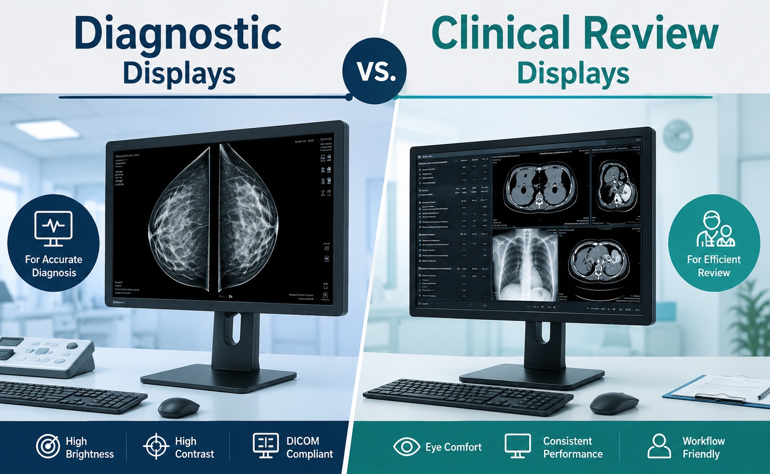
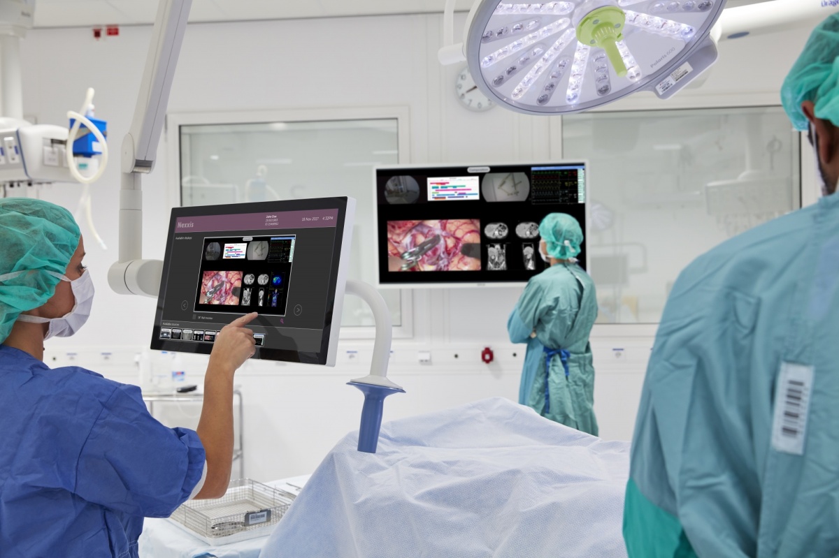

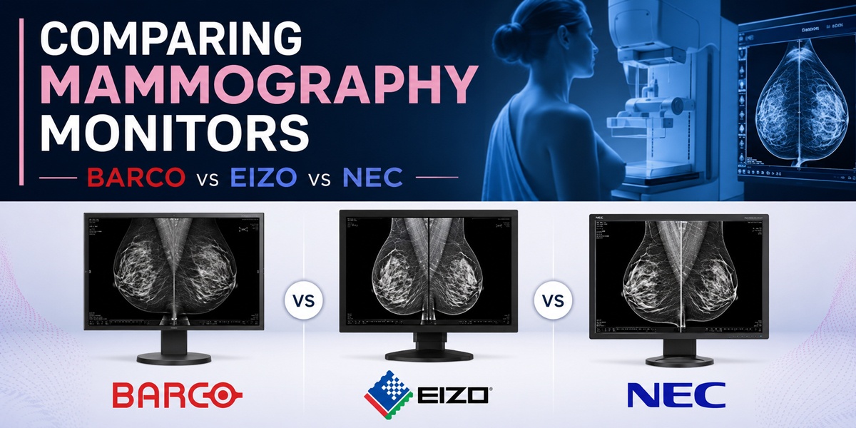
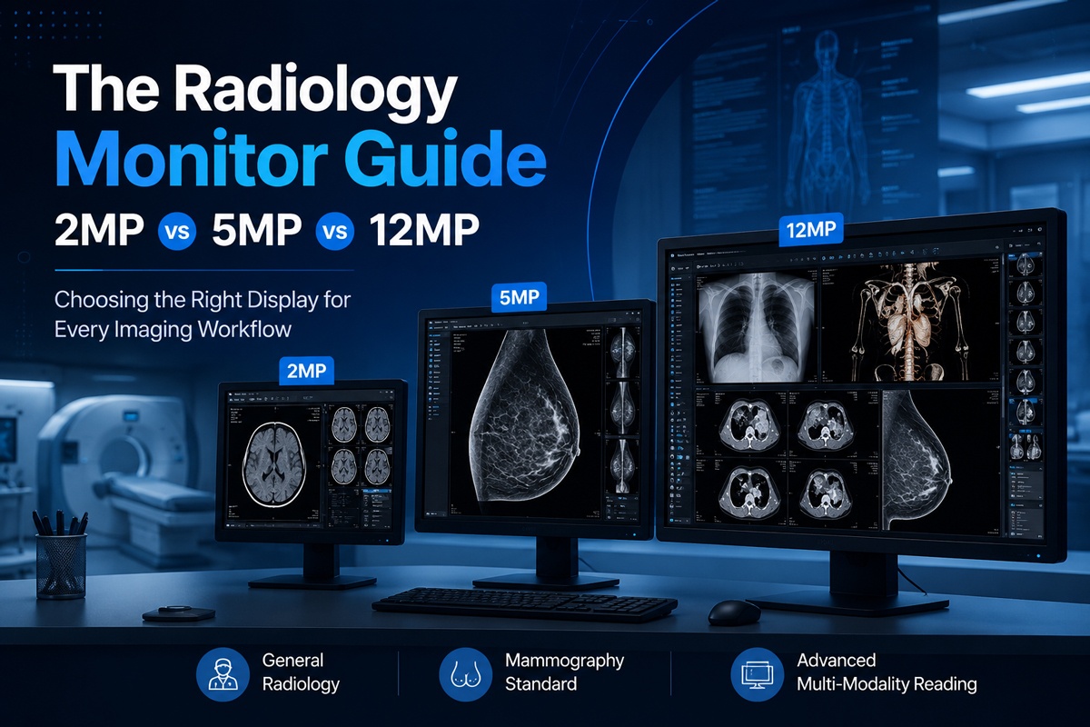
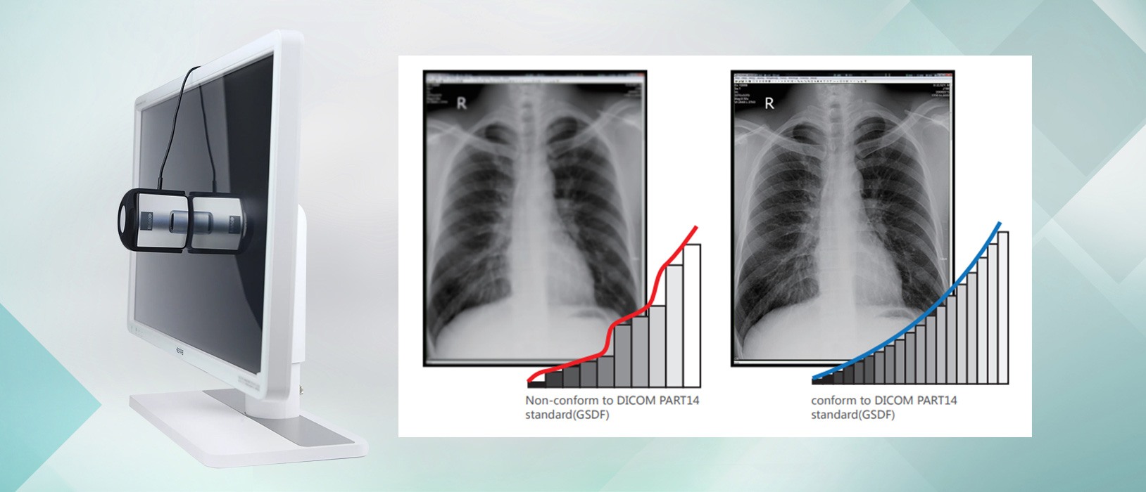
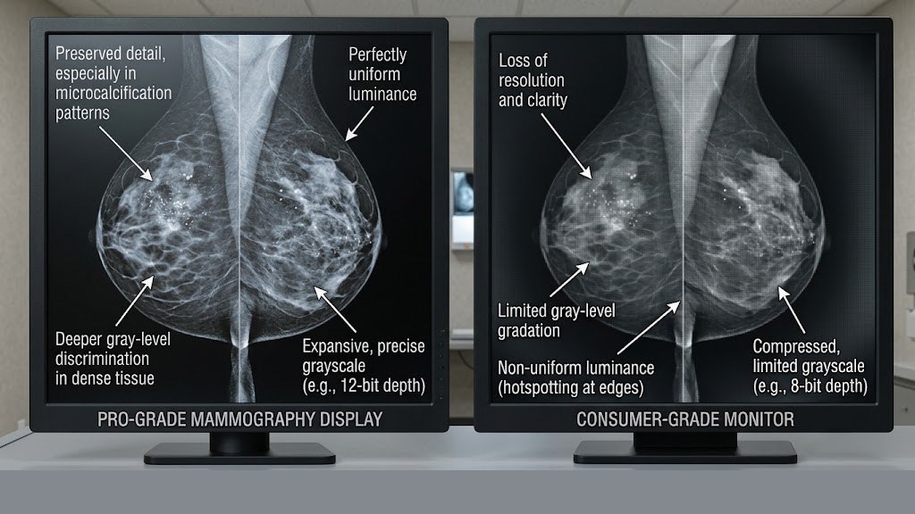
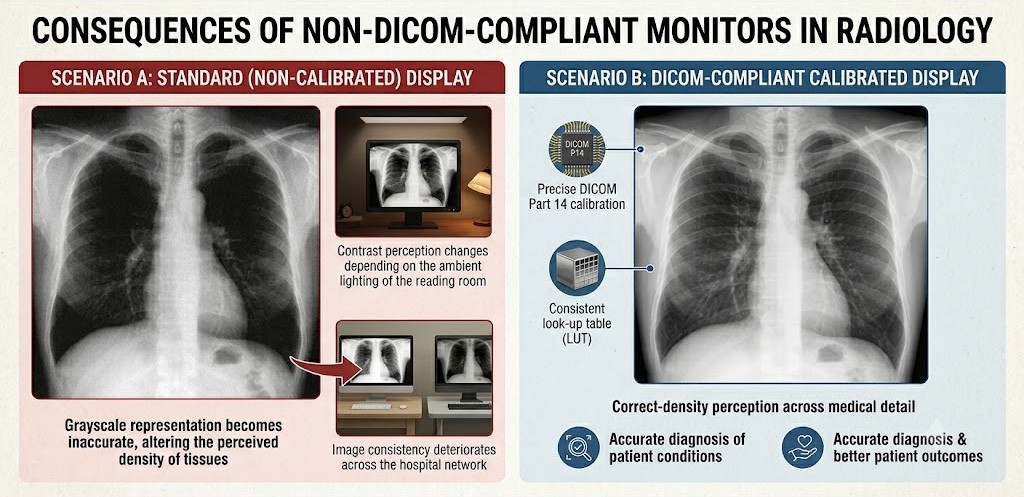

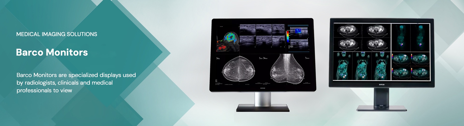